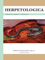
This paper represents the first ultrastructural descriptions of the cloacal glands of a male salamander. All male salamanders in the suborder Salamandroidea possess these glands, which are required for internal fertilization. These glands are responsible for producing spermatophores and, in some species, certain glands produce female-attracting pheromones. Both scanning and transmission electron microscopy provided higher resolution and magnification of the cytology of cloacal glands than possible in previous studies limited to light microscopy. The glands are hypertrophied in October and April samples, and much reduced in size and secretory activity in June and August samples. Each of the cloacal glands, when active, has secretory vacuoles that are unique in appearance and that secrete carbohydrates and/or proteins. Kingsbury's glands are modified mucous glands that have secretory vacuoles filled with a flocculent substance. Active pelvic glands have uniform electron-lucent secretory vacuoles that fill the cytoplasm and narrow the lumen. Ventral glands have squamous epithelium with moderately dense vacuoles scattered around the apical border, and a granular secretion that fills the wide lumen. In the breeding season, vent glands have biphasic secretory vacuoles characteristic of glycoproteins. All glands release their products by a merocrine process, and all have myoepithelial sheaths, which are best developed in ventral glands. Future research should focus on differences in cytology among the pheromone producing glands and comparisons with the ventral glands in salamander clades that do not produce spermatophores (e.g., Hynobiidae and Cryptobranchidae).
How to translate text using browser tools
1 March 2014
Ultrastructure of the Male Cloacal Glands of the Eastern Red-backed Salamander, Plethodon cinereus
David M. Sever
ACCESS THE FULL ARTICLE

Herpetologica
Vol. 70 • No. 1
March 2014
Vol. 70 • No. 1
March 2014
Glandular activity
microanatomy
Pheromone glands
Reproductive histology
Spermatophore glands


