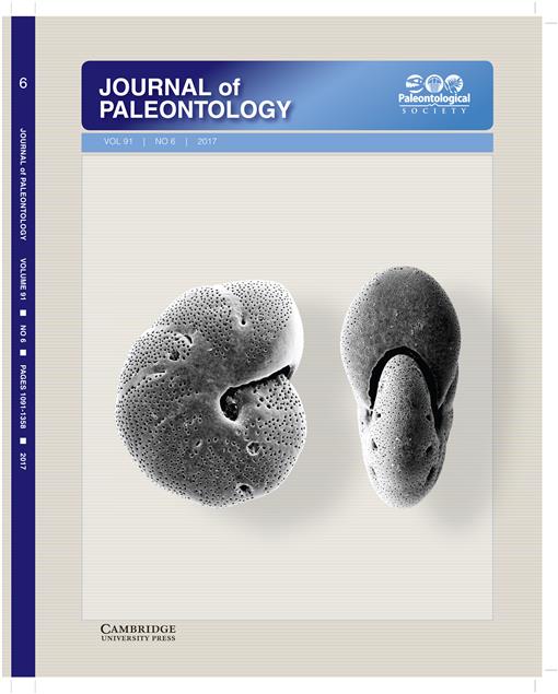
Earliest Triassic natural conodont assemblages preserved as impressions on bedding planes occur in a claystone of the Hashikadani Formation, which is part of the Mino Terrane, a Jurassic accretionary complex in Japan. In this study, the apparatus of Hindeodus parvus (Kozur and Pjatakova, 1976) is reconstructed using synchrotron radiation micro-tomography (SR—μCT). This species has six kinds of elements disposed in 15 positions forming the conodont apparatus. Carminiscaphate, angulate, and makellate forms are settled in pairs in the P1, P2, and M positions, respectively. The single alate element is correlated with the S0 position. The S array is a cluster of eight ramiforms, subdivided into two inner pairs of digyrate S1–2 and two outer pairs of bipennate S3–4 elements. The reconstruction is similar to a well-known ozarkodinid apparatus model. In addition, the μCT images show that the ‘anterior’ and ‘posterior’ processes of the S1–2 elements faced the caudal and rostral ends of the living conodont body, respectively.
How to translate text using browser tools
1 November 2017
Reconstruction of the multielement apparatus of the earliest Triassic conodont, Hindeodus parvus, using synchrotron radiation X-ray micro-tomography
Sachiko Agematsu,
Kentaro Uesugi,
Hiroyoshi Sano,
Katsuo Sashida
ACCESS THE FULL ARTICLE

Journal of Paleontology
Vol. 91 • No. 6
October 2017
Vol. 91 • No. 6
October 2017


