Here we describe three new notohippid notoungulate species from the early Oligocene-aged Tinguiririca Fauna (Tinguirirican SALMA), recovered from volcaniclastic deposits of the Abanico Formation in the central Chilean Andes, two of which are known from material sufficiently complete to warrant formal naming. These include Eomorphippus bondi, sp. nov., a form of moderate size distinguished by hypsodont incisors and cheekteeth, as well as distinctive proportions of the upper incisors. A closely similar but more diminutive form is described as Eomorphippus neilopdykei, sp. nov. A third previously unrecognized notohippid in the Tinguiririca Fauna, best represented by a large, low-crowned, lower incisor battery, almost certainly represents a new taxon, but remains too fragmentary to warrant naming now. We also propose a new binomial for a previously named notohippid, ?Eomorphippus pascuali, originally described from Gran Barranca in Argentina but which is now also recorded in Chile. This taxon, here named Rosendo pascuali, is markedly less hypsodont than E. bondi and E. neilopdykei and retains lingual cingula on at least p4-m1. As least one leontiniid notoungulate occurs in the Tinguiririca Fauna, Termastherium flacoensis, gen. et sp. nov., best represented by two partial upper cheek toothrows and a tentatively referred maxillary fragment bearing three deciduous teeth. Collectively, description of these new fossils from Termas del Flaco, Chile helps to more fully characterize the Tinguiririca Fauna, facilitating correlation and comparison to other South American land mammal faunas spanning the Eocene-Oligocene transition.
INTRODUCTION
Only in the past two decades has it been realized that the widespread volcaniclastic deposits of the Andes of central Chile host one of the most important archives of Cenozoic mammalian evolution in South America. The first mammalian fauna discovered in this region, the Tinguiririca Fauna of the upper Río Tinguiririca drainage (Novacek et al., 1989), has since been designated the type fauna of the Tinguirirican South American Land Mammal “Age” (SALMA) (Flynn et al., 2003). The Tinguirirican is currently regarded as temporally interposed between the Mustersan (late Eocene) and Deseadan (late Oligocene—early Miocene) intervals of the classical SALMA sequence, given that the Divisaderan, originally considered to precede the Tinguirirican, has recently been shown to be invalid as it represented a mixed assemblage of demonstrably older (mainly Casamayoran) and younger (Deseadan to Santacrucian) taxa (Cerdeño et al., 2008; López, 2008, 2010; López and Manassero, 2008). Following a preliminary accounting (Wyss et al., 1994), various components of the Tinguiririca Fauna have been described in detail elsewhere, including its marsupials (Flynn and Wyss, 1999), interatheriids (Hitz et al., 2000, 2006), archaeohyracids (Croft et al., 2003; Reguero et al., 2003), tardigrades (McKenna et al., 2006), dasypodids (Carlini et al., 2009), rodents (Bertrand et al., 2012), and notostylopids and basal toxodontians (Bradham et al., 2015). Here we build on this documentation, describing the faunas diverse but sparsely represented notohippids and leontiniids.
A spate of studies over the past two decades has enhanced understanding of the taxonomy and phylogeny of notohippid notoungulates. Noteworthy among these has been the description of one of the earliest known representatives of the clade, Pampahippus arenalesi from the Lumbrera Formation of northwestern Argentina, and referral of several Casamayoran and Mustersan taxa from Patagonia, traditionally regarded as isotemnids, to the Notohippidae (Bond and López, 1993). Additional species of Pampahippus have been described, P. secundus (Deraco and García-Lopez, 2016) and P. powelli (García-López et al, 2017). Shockey (1997) described three notohippids based on superbly preserved material from the Deseadan of Bolivia, providing the first cladistic parsimony analysis of relationships within the group. Gabbert (2004) described the basicranial anatomy of the Mustersan taxon, Puelia, contributing data relevant to unraveling higher-level toxodontian interrelationships. A new late Oligocene notohippid from Quebrada Fiera (Mendoza, Argentina) was described in the context of a phylogenetic analysis of the more inclusive group to which it belongs (Cerdeño and Vera, 2010,2014). López et al. (2010) documented notohippids from the “La Cancha” level (also referred to as a fauna and a locality) at the Gran Barranca in Patagonia, apparently Tinguirirican in age, recognizing a large number of species (five) from that level.
The names of the major subgroups of notoungulate mammals to which the species described below belong are not currently employed consistently. This nomenclatural instability has two primary sources. First, the continued application of traditional Linnaean ranks and differences of opinion about which rank each group warrants have led to interchangeable endings of some names. Second, continuing uncertainty remains about which of the historically recognized notoungulate subgroupings constitute monophyletic entities, and thus warrant naming in the first place. Unfortunately, both of the species described below fall within one of the nomenclaturally most challenged branches of the notoungulate evolutionary tree. Of the four widely recognized notoungulate “subordinal,” groupings, the species described below are members of what is variously termed the Toxodonta (Scott, 1905) or Toxodontia (Owen, 1853); see conflicting usages in, e.g., Simpson (1967), Mones (1986), and McKenna and Bell (1997). This group as a whole has yet to be diagnosed rigorously, and two of its five traditionally recognized subdivisions are demonstrably nonmonophyletic: Isotemnidae and Notohippidae (in contrast to Homalodotheriidae, Leontiniidae, and Toxodontidae, each of which clearly are monophyletic).
Although differing in some details, the analyses of Cifelli (1993), Shockey (1997), Cerdeño and Vera (2010), Billet (2011), and Shockey et al. (2012) have shown that the clade encompassing the species conventionally regarded as notohippids is nonexclusive, that is, the most recent common ancestor (MRCA) shared by these species also gave rise to toxodontids and, potentially, leontiniids. Bond and López's (1993) recognition of the notohippid affinities of various Casamayoran and Mustersan species formerly regarded as isotemnids and the subsequent questioning of these results (Cerdeño and Vera, 2010) only underscore the need to stabilize toxodontian taxonomy. The name Notohippidae, in particular, need not remain in such a confused state (referring simultaneously to differing sets of taxa). An obvious remedy would be to apply a phylogenetic definition, whereby some stem, node, or apomorphy would specify the clade to which the name is unambiguously linked. Formally proposing a phylogenetic definition for the name Notohippidae lies beyond the scope of the present analysis, however, given the poorly resolved understanding of relationships among notoungulates in general and notohippids in particular. Nevertheless, we urge that Notohippidae ultimately be defined phylogenetically. In the interim, we employ the name Notohippidae informally, to refer to a monophyletic entity that approximately encompasses the MRCA of Pampahippus and some particularly well-known, later-diverging member of the clade such as Rhynchippus or Notohippus, plus all its descendants. Although with our current understanding of phylogenetic relationships this would make the Toxodontidae and Leontiniidae members of a clade bearing a name with an identical ending (-idae), such a circumstance is easily accommodated within a phylogenetic system. Alternatively, in a more traditional Linnaean classification, a higher-level clade name could be created for this group.
Future analyses might also demonstrate homalodotheriids to be members of the Notohippidae (as the latter name is employed here), raising the question of whether the clade just described might more appropriately be named Toxodontia (in which case, the present report would more appropriately have been titled, “New Paleogene Toxodontians from…”). Nevertheless, we have adopted the usage indicated above to avoid confusion with other uses of the name Notohippidae and to meet our immediate need to succinctly refer to the clade encompassing the species described below plus their nearest allies. We hope that this stopgap measure will be temporary, inasmuch as definitive phylogenetic definitions of this name and those associated with other clades of extinct South American ungulates are long overdue.
ABBREVIATIONS
Institutional abbreviations for specimens referred to in this study are: AMNH FM, American Museum of Natural History, Division of Paleontology Fossil Mammal Collections; FMNH, Field Museum of Natural History; ICN-P, Instituto de Ciencias Naturales y Museo de Historia Natural, Universidad Nacional de Colombia, Bogotá; MLP, Museo de La Plata; and SGOPV, Museo Nacional de Historia Natural, Santiago. Dental abbreviations: L, left; R, right; i/I, lower/ upper incisor; c/C, lower/upper canine; p/P, lower/upper premolar; dp/P, deciduous lower/ upper premolar; and m/M, lower/upper molar.
SYSTEMATICS
Notoungulata Roth, 1903
Toxodontia Owen, 1853
Notohippidae Ameghino, 1894
Eomorphippus Ameghino, 1901
Type Species: Eomorphippus obscurus Ameghino, 1901.
Diagnosis (emended from Simpson, 1967): Marked hypsodonty of the upper and lower incisors, posterior premolars, and molars. Cementum lacking. Upper incisors moderately procumbent. I3 broad relative to I2 and I1. P4 more molariform than the anterior premolars, but lacking a distinct hypocone. Upper molars bearing hypocones, with a variable but deep cleft separating them from the protocone, the cleft blocked by the medial projection of what is likely the anterior end of the crochet. Other than the persistent major fossa, fossettes are obliterated early in wear.
Simpson (1967) also listed P1–3 with notched or incomplete protolophs as diagnostic of Eomorphippus, but this feature applies to notohippids in general (Bond and López, 1993). He also considered lower molars with clefts anterior and posterior of the entoconid that variably develop into short-lived fossettids with wear as diagnostic of Eomorphippus, but this feature also appears to characterize a more inclusive group.
Eomorphippus bondi, species novum
Figure 1
Holotype: SGOPV 3046: partial skull preserving orbital rims, portions of both zygomatic arches, rostrum, and LI1–C, P3–M3; RI1–C, P3, M1–3.
Paratype: SGOPV 2891, partial left lower dentition including i1–3 and p2–m3, plus remnants of the right dentition, including i1 and molds and external (labial) slivers of some posterior cheekteeth.
Tentatively Referred Specimen: SGOPV 3085, labiolingually crushed right ?p4–m1.
Type Locality: The type, paratype, and tentatively referred specimen all derive from volcaniclastic sediments currently mapped as belonging to the Abanico (= Coya-Machalí) Formation in the Tinguiririca River valley (∼35° S), in the Andean Main Range of central Chile, some 7 km west of the international boundary. (Geologic maps dating from before the late 1980s mistakenly identified these deposits as belonging to the Cretaceous Colimapu Formation; Charrier et al., 1996.) All specimens described below were recovered from 35°–50° west-dipping strata, north of an unnamed 2738 m pass (indicated on the topographic sheet; Anonymous, 1985), approximately 3 km south of the town of Termas del Flaco, at what is termed the “main locality” in Charrier et al. (1996: fig. 6).
Age: Early Oligocene (to potentially late Eocene), Tinguirirican SALMA. The diverse Tinguiririca Fauna recovered from near Termas del Flaco, of which the notohippids are an important constituent, formed the basis for the formalized Tinguirirican SALMA (Flynn et al., 2003), which lies temporally between the Mustersan and Deseadan of the classical SALMA sequence. An extensive series of isotopic dates (summarized in Flynn et al., 2003) have been generated for strata hosting and underlying the Tinguiririca Fauna, indicating that E. bondi and its contemporaries described below are no younger than ∼31.5 million years old (early Oligocene) and could be ∼1–2 million years older (Bradham et al., 2015).
Etymology: In honor of Mariano Bond, for his enormous and influential contributions to the understanding of notoungulate phylogenetics and taxonomy.
Diagnosis: Eomorphippus bondi generally resembles E. obscurus, differing from the latter mainly in being roughly 20% larger in most dental dimensions, in having a slightly smaller upper canine, and in having an I3 that is substantially wider than both I1 and I2.
DESCRIPTION
Eomorphippus bondi is the best-represented notohippid from the Tinguiririca Fauna and one of two relatively large-bodied species from the assemblage. The holotype has previously been referred to as Eomorphippus n. sp. (Wyss et al., 1994) and Eomorphippus undesc. sp., near E. obscurus (Flynn et al., 2003). Two specimens from Patagonia are of particular importance to our comparisons and identification of the new Chilean material. The first is MLP 12-1508, the holotype of Eurystomus stehlini Roth, 1901, collected by Santiago Roth from the enigmatic locality Cañadón Blanco (see below). This specimen consists of most of the upper and lower dentition and was referred by Simpson (1967), on Bryan Pattersons recommendation, to Eomorphippus obscurus Ameghino, 1901. The second specimen is a nearly complete upper dentition that Egidio Feruglio collected from the Gran Barranca and deposited in the collections of the University of Padua, Italy. Simpson (1967) referred this specimen to E. obscurus, a cast of which is deposited at the AMNH (AMNH FM 27885).
Upper Dentition: A partial skull, SGOPV 3046, preserves a nearly complete upper dentition, serving as the sole record of the upper dentition of this taxon. This specimen has been slightly distorted through lateral compression, thereby slightly exaggerating the closeness of the posterior portions of the opposing toothrows, and possibly reducing the transverse dimensions of the cheekteeth, particularly the molars. Measurements are given in table 1.
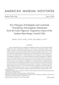
FIGURE 1.
Photographs of cast, and line drawings of holotype of Eomorphippus bondi, SGOPV 3046, a partial skull bearing left I1–3, C, P3–M3 and right I1–3, P3, M1–3, in A, left lateral, B, C, occlusal, and D, anterior views (opposite page). Horizontal ridge in anterior view is a seam from the two-piece mold. Note hypsodonty of the incisors and narrowness of molars. Paratype of Eomorphippus bondi, SGOPV 2891, showing partial left lower dentition including i1–3 and p2–m3, in E, occlusal and F, labial views (above; photograph of cast).

Continued.
Photographs of cast, and line drawings of holotype of Eomorphippus bondi, SGOPV 3046, a partial skull bearing left I1–3, C, P3–M3 and right I1–3, P3, M1–3, in A, left lateral, B, C, occlusal, and D, anterior views (opposite page). Horizontal ridge in anterior view is a seam from the two-piece mold. Note hypsodonty of the incisors and narrowness of molars. Paratype of Eomorphippus bondi, SGOPV 2891, showing partial left lower dentition including i1–3 and p2–m3, in E, occlusal and F, labial views (above; photograph of cast).

The three upper incisors form an impressive cropping apparatus arranged in a smooth arc. They are moderately procumbent, as is also well seen in AMNH FM 27885 (the Feruglio specimen of E. obscurus; see Simpson, 1967: fig. 38). Lingual cingula (as have been noted for E. obscurus by Patterson in Simpson, 1967) appear to be absent, but this may simply reflect a fair degree of wear on I1–2 in SGOPV 3046. Rectangular wear facets occur on the lingual faces of the four central incisors. The centrally placed, blunt ridge present on the lingual face of I3 in E. obscurus (Patterson in Simpson, 1967) does not occur in E. bondi; instead, a broadly concave wear facet spans much of this portion of the tooth. All three incisors are moderately curved posteriorly along their substantial height. These teeth are faced anteriorly with broad, featureless sheets of enamel. Breakage of the distalmost surficial layer of the premaxilla exposes much of the proximodistal height of these teeth, including large portions that were undoubtedly well within the alveolus (well above the gumline) in life. Significantly, enamel reaches nearly (if not completely) to the tips of the roots of these teeth (fig. 1 A, D). This remarkable degree of incisor hypsodonty is best seen on RI3, where the posterior margin of the tooth is exposed nearly to the tip of its root. The three incisors become progressively broader transversely, the mesiodistal diameter of I3 being roughly twice that of I1 (table 1, fig. 1D). The muzzle is broadest across the distal portions of the I3s, becoming progressively more constricted until roughly the anterior margin of P3, at which point the palate gradually widens again.
The upper canine, preserved only on the left side, is greatly reduced in size in comparison to the incisors. Seemingly unworn, this tooth may not yet be fully erupted, as its tip is ~1 cm more dorsal than the bladelike termination of I3, although a similar relationship between the height of the canine and I3 is exhibited by AMNH FM 27885. The tooth is strongly canted anteriorly and situated posterior and slightly medial to the posterolateral margin of I3. The alveolus for a tooth of similar size and position is preserved on the specimen's right side. The canine of SGOPV 3046 is just a sliver of a tooth compared to the incisors, much smaller than that of MLP 12-1508 and more similar to that of AMNH FM 27885—both of which Simpson (1967) referred to E. obscurus.
Neither P1 nor P2 is preserved. A faint outline of an alveolus occurs midway in the gap between the canine and P3 on the left side of the specimen. Judging from the length of this gap and the size of the posterior premolars, no more than small spaces likely separated the teeth between the canine and the third premolar. A largely closed upper toothrow is also present in E. obscurus (Simpson, 1967).
Both P3s are present and subtriangular in outline. It and all succeeding cheekteeth are moderately bowed. A strong paracone column or fold marks the high labial face of the tooth anteriorly. This structure, together with the small parastyle emanating from it, forms a strong anterolabial pillar. On the specimens left side, the posterior half of the labial face of this tooth is overlain by the parastyle of P4. This region of the tooth is exposed on the right side, since RP4 is missing, showing a subdued metacone fold. The ectoloph projects far beyond the rest of the occlusal surface of P3, which includes a large central fossa and a protocone forming its lingual border. Wear has obscured details of the loph arrangement, and evidence of any cingulum is lacking, in contrast with the condition in E. obscurus (Simpson, 1967).
The P4 is more trapezoidal in outline than P3, with the tooth's lingual side consisting of a broad, flat face. The tooth's labial surface is basically a larger version of the arrangement seen on the preceding tooth (P3), except for its more prominent parastyle. The protoloph is complete and reaches the ectoloph anterolabially to form the flat lingual face of the tooth. The enamel near the lingual end of the posterior face extends slightly into the occlusal surface, and a minute fossette on the posterior crown surface suggests that a posterior cingulum may have been present earlier in wear. The anterior face of the tooth shows no evidence of a cingulum, again in contrast to the condition in E. obscurus (Simpson, 1967).
The first two molars are quite similar in form, differing mainly in size and degree of anteroposterior elongation. All three molars are mildly imbricated. The paracone and metacone folds on the labial faces of the molars are less pronounced than in P4, but the molars all retain a short parastyle. Given the height of these teeth, their cross-sectional shape and dimensions would have changed substantially during wear. Beginning as mesiodistally elongate π-shaped structures, the exposed bases of the crowns indicate that a much squarer occlusal surface outline would have been achieved later in life. A strong cleft divides the protocone and the hypocone columns lingually. On M1, the protocone column makes up roughly two-thirds of the length of the concave lingual face of the tooth at the wear surface; on M2 the cleft is wider than on Ml at the level of the occlusal surface (narrowing dorsally toward the crown base). A small fossa on the hypocone column of Ml and M2 presumably partly represents the remnants of a posterior cingulum. M3 is incompletely erupted and little more than the anterior portion of its labial face and protocone column are visible.
Lower Dentition: Most elements of the lower dentition are represented in SGOPV 2891 (fig. 1E, F). This specimen consists of a pair of toothrows, the left side of which is largely complete. The right side preserves only slivers and natural molds of the labial faces of the posterior cheekteeth and a poorly preserved i1. SGOPV 2891 is referred to E. bondi based on its compatibility in size and morphology to the upper dentition of the holotype.
Of the incisor battery, Ril and Li1–3 are preserved but are somewhat damaged and not cleanly separated from the matrix. These teeth appear to be slightly dislocated ventrally with respect to the remainder of the toothrow, but since no trace of the mandibular ramus is preserved, this is difficult to ascertain with confidence. All three incisors are spatulate in form, perhaps increasing slightly in mesiodistal breadth posteriorly. The lingual faces of these teeth remain covered in matrix, obscuring whether the conspicuous ridges seen in E. obscurus are present. The presence of wear facets is masked by poor preservation. The lower incisors are much more feebly developed than the uppers, the former measuring at most half the width of the latter. The lowers are arranged in a much tighter arc than their upper counterparts. Although the lower incisors are far less hypsodont than the uppers (the left i2, exposed clearly to the tip of its root, measures ~15 mm in height, while the right I2, which is incompletely exposed, measures >23 mm), enamel can be seen to cover not just the crowns, but also at least the labial faces of the roots. The lower incisor arcade exhibits none of the reduction in number or pattern of enlargement indicative of leontiniid affinities. Moreover, it is considerably narrower than the broad upper incisor/canine complex. This structural asymmetry between the upper and lower incisors is curious. In other respects, this lower dentition has the morphology and size expected for this species, and we have not recovered any lower dental elements in the Tinguiririca Fauna assemblage that would be better candidates for referral to E. bondi.
A transversely oriented, crescentic sliver of enamel is present in a short, matrix-filled gap posterior to Li3; this probably represents a damaged remnant of p1, or a dislocated fragment of the canine. The remains of this tooth indicate that the diastema between the incisor series and the next most posterior tooth was no more than 5 mm long, and thus that the toothrow was largely closed. If a canine once was present, no clear evidence of it is preserved.
The three posterior premolars, all little worn, increase in size posteriorly. A large, sharply angular trigonid dominates the crown of p2. The mesial end of the trigonid is slightly damaged, but a short, mesially projecting protolophid appears to have been present. There is no evidence of a paralophid. The metalophid is broad labiolingually, its apex forming a crest that is essentially transverse but mildly concave posteriorly. The lingual end of this crest is continuous posteriorly with a prominent cristid obliqua that delimits a feeble, open talonid labially. In labial view the anterior end of the cristid obliqua slopes posteriorly; the posterior face of the trigonid slopes even more steeply posteriorly (∼70°). The cristid obliqua is set quite far lingually, the entire length of the hypolophid occurring within the medial half of the tooth. Anteriorly it joins the paraconid rather than the center of the metalophid. There is no evidence of an entoconid.
TABLE 1.
Mensural data for specimens described in text. Asterisk denotes uncertainty due to damage of the specimen in question.

Continued

The trigonid of p3 is similar to that of p2 in being dominated by a transverse, posteriorly concave metalophid, which projects labially as a strong protoconid ridge. As on p2, this ridge is canted anteriorly, and forms the farthest-projecting portion of the tooth labially. A protolophid extends anterolingually from the point where the labial extremity of the metalophid plunges into the protoconid ridge. The protolophid descends to align almost perfectly with the posterior terminus of the p2 hypolophid. Although p2–3 are tightly appressed, no paralophid appears to have been present on p3. Seen in occlusal view, the two trigonid lophids resemble the seven symbol (i.e., 7), the short arm of which represents the protoconid ridge. As on p2, the p3 hypolophid is medially positioned and contacts the metaconid anteriorly; it is considerably shorter mesiodistally than the trigonid. The hypolophid is more convex lingually than on p2; within this hollow sits a low, broad entoconid, which is separated from the trigonid anteriorly by a deep sulcus. The lingual face of p3 is well preserved and cleanly exposed to its base. Remarkably, a strong, ~1 mm wide, lipped cingulum rims the base of p3 lingually; it is slightly papillate and runs the length of the tooth very low on the crown. It climbs at both its posterior and anterior ends to join the vertical postero- and anterolingual edges of the talonid and trigonid respectively. Such a strong cingulum is unusual for post-Mustersan notohippids. Preservation of the lingual faces of p2 and p4 is too poor to establish whether cingula were present.
Although slightly larger than p3, the posteriormost premolar resembles the anterior premolars more than the molars. The cleft separating the trigonid and talonid labially is shallower than on p2–3, but deeper than on the molars and slightly more vertical. The p4 trigonid resembles that of p3; there is no evidence of a paralophid and the protolophid projects directly into, and is appressed against, the posterior end of the hypolophid of p3. The short hypolophid is not well preserved posteriorly, but it attaches to the posterior trigonid wall lingually. Thus, on p4 the cristid obliqua meets the metaconid, while on the molars it meets the metalophid more centrally.
The complete heights of the lower premolar crowns are preserved and exposed labially. A faint cingulum descends from below the anterior extremity of the protolophid of p2, traversing the base of the trigonid and protoconid column. It encloses a small basin at the base of the corner formed between the lingually offset hypolophid and the trigonid/talonid cleft. A barely perceptible cingulum slants across the base of the p3 trigonid labially, while none is evident on p4.
One somewhat enigmatic specimen, SGOPV 3085, is tentatively referred to E. bondi. It consists of two laterally crushed lower cheekteeth, probably p4–m1, both of which are noteworthy for bearing strong lingual cingula. The lingual faces of these teeth are completely intact, but their labial halves are largely imploded. The uncrushed paralophid of the anterior tooth, and the continuous, sweeping ectoloph on the posterior tooth mark these teeth as belonging to the right dental series. To the degree that this specimen can be compared to the lower dentition of the paratype, the agreement in size and morphology are quite close. The cingulum is especially long and strong on the anterior tooth, closely resembling the cingular configuration of p3 on SGOPV 2891. It forms a broad, low, U-shaped gutter, swinging dorsally at its anterior end (where it merges with the paralophid) and at its posterior end (where it rises to the hypoconulid). Its crest is generally smooth, bearing few, if any, papillae. The posterior tooth (questionably identified as m1) bears a similarly strong lingual cingulum across the anterior half of its base that extends just slightly posterior to the trigonid/talonid junction. These lingual cingula are more pronounced than in SGOPV 2996 (referred to the Leontiniidae below), but this latter specimen consists of a more posterior portion of the dentition (where the cingula might be expected to be weaker).
The molars are of quintessential notoungulate form, consisting of a short, strongly lophate trigonid, and a much longer, equally highly lophate talonid. The paraconids and metaconids are separated by a deep, lingually sloping, bowl-shaped cleft. This excavation is obliterated early in wear, resulting in a featureless occlusal wear surface. The talonid is dominated by a long hypolophid, which forms a smooth, labially convex surface making up the posterior two-thirds of the labial faces of the molars. Lingual curvature of the posterior end of the hypolophid is least pronounced on m3, which has the proportionally longest talonid. With wear, the entoconid forms a broad lophid continuous with the hypolophid labially. Clefts initially separate the entolophid from the trigonid anteriorly and from the “tail” of the hypolophid posteriorly. These clefts close progressively with wear, the anterior one more rapidly. On m1 the anterior cleft has been reduced to a small fossettid at the trigonid/talonid juncture, while on m2 the anterior cleft persists as a transverse slit. The m2 is the only molar on which the lingual face is reasonably well preserved; a cingulum highly reminiscent of the one seen on the posterior tooth of SGOPV 3065 spans the base of the trigonid approximately 7 mm below the occlusal surface. The m3 is not fully erupted, obscuring details of this tooth's posterolingual quadrant, which remains buried in the matrix-filled crypt.
Based on the presence of lingual cingula, SGOPV 2891 might seem better referred to the Leontiniidae (see below) rather than the Notohippidae, as we have done here. The lack of a caniniform or tusklike i3 in SGOPV 2891, a feature diagnostic of leontiniids, strongly argues against referral to that group, however. It is thus difficult to escape the conclusion that two Tinguirirican taxa, R. pascuali, and E. bondi, are unusual among post-Casamayoran notohippids in retaining lingual cingula on at least some lower cheekteeth.
Age Implications: Eomorphippus is classically regarded as Mustersan in age (Simpson, 1967; Marshall et al., 1983). Discovery of the biochronologically and radioisotopically welldelimited Tinguiririca Fauna and the ramifications this had for interpreting roughly contemporaneous assemblages from Patagonia have led to a reassessment of this view, indicating that Eomorphippus does not occur earlier than the latest Mustersan, if it occurs in the Mustersan at all (López et al., 2010).
E. obscurus is best known from the enigmatic Cañadón Blanco Fauna collected early in the 20th century by Santiago Roth; for decades thereafter this fauna was considered to represent a chronologically mixed assemblage, including specimens of presumed Deseadan, Mustersan, and Casamayoran age. There was no firm basis, however, for judging assignment of taxa from Cañadón Blanco to the specific SALMA it supposedly represented. Nevertheless, the record of Eomorphippus from Cañadón Blanco became the primary basis for regarding this taxon as characteristic of the Mustersan. In view of the suite of taxa cooccurring in the temporally cohesive Tinguiririca Fauna of Chile, most of the specimens from Cañadón Blanco are now regarded as pertaining to a single fauna, one roughly coeval with that Chilean fauna (Wyss et al., 1994).
E. obscurus has been reported from elsewhere in Patagonia, notably from the Gran Barranca south of Lake Colhué-Huapí, but here again evidence counters the assumed “typical” Mustersan age of some of these occurrences. First, some specimens collected from the Gran Barranca by C. Ameghino are labeled as from the “Partie la plus supérieure de couches à Astraponotus” (latest Mustersan SALMA in current terminology), or “APS” (Astraponotus plus supérieure [or “Astraponotéen plus supérieur”]) as termed by Bond et al. (1996, 1997) and Kay et al. (1999). Second, Simpson (1967) tentatively referred a specimen collected by his team at the Gran Barrranca (AMNH FM 29462) to E. obscurus, making particular mention of its recovery from high within his Mustersan section. Similarly, an upper dentition, the most complete specimen of E. obscurus known from the Gran Barranca, discovered by Egidio Feruglio and described by Simpson (1936) originally as Pseudostylops subquadratus, was collected from the upper part of Feruglio's level 3f, a 14 m thick, “concretionary, corniceforming tuff, so-called ‘tosquilla’” (Simpson, 1936: 5). Simpson regarded the entirety of level 3f to be Mustersan in age, viewing the Casamayoran-Mustersan transition as occurring at the contact between level 3f and the underlying 3e or possibly near the top of 3e. Significantly, the horizon producing E. obscurus was the highest fossiliferous level in Feruglio's section, meaning that this specimen likely originated from a stratigraphic level chronologically equivalent to Ameghino's APS (presumably Ameghino and Feruglio's sampling localities at the Gran Barranca did not coincide exactly). Three different collectors (Ameghino, Feruglio, and Simpson) thus independently recovered E. obscurus from the highest levels of beds once considered Mustersan, but that are now regarded as pertaining to the Tinguirirican SALMA (Wyss et al., 1994; Bond et al., 1996, 1997; Kay et al., 1999; Flynn et al., 2003; Gelfo et al., 2009). This age inference is supported by recent studies at Gran Barranca that have documented Eomorphippus only at level GBV-4 (“La Cancha”), an interval referred to the Tinguirician SALMA (López et al., 2010) (see also: Ré et al., 2010; and Dunn et al., 2013). Moreover, well-sampled deposits of undisputed Mustersan age in Patagonia have not yielded E. obscurus, despite extensive collecting efforts there, and the occurrence of other notohippid species at these localities (M. Bond, personal commun.). Lastly, E. obscurus has not been identified in preliminary analyses of pre-Tinguirirican faunal assemblages from the richly fossiliferous Abanico Formation assemblages elsewhere in Central Chile. In sum, it now appears that Eomorphippus obscurus (and the closely related E. bondi, as well as the new species E. neilopdykei; see below) are restricted to the Tinguirirican SALMA.
Eomorphippus neilopdykei, species novum
Figure 2
Holotype: SGOPV 2855, pair of mandibles preserving at least one representative of each lower tooth locus except the canine and p1.
Paratype: SGOPV 3071, a right mandible bearing five or more teeth, apparently m2 and more anterior loci, but only partially prepared.
Type Locality: As for Eomorphippus bondi.
Age: As for Eomorphippus bondi.
Etymology: In recognition of Neil Opdyke, for his pioneering research on paleoclimate and plate tectonics, and career-long support of integrative studies of vertebrate paleontology and geology. He fondly recalls serving as a “pack animal” while he was an undergraduate field assistant for Keith Runcorn, making it especially appropriate to name a fossil evoking a “beast of burden” in his honor.
Diagnosis: As for E. obscurus and E. bondi, but differing from those species mainly in much smaller size.
Description: Eomorphippus neilopdykei, identified as “notohippid new taxon B” by Wyss et al. (1994) and as “undescribed taxon B” by Flynn et al. (2003), is currently known only from lower teeth. E. neilopdykei is nearly identical in all comparable morphological details to E. bondi, except that it is much smaller at only slightly greater than half the size of the latter species.
Lower Dentition: The partial rami of the fused pair of mandibles making up the holotype have been mildly deformed by tectonic compression, but the teeth are uncracked and otherwise show no evidence of distortion. The left ramus is broken ventral to p2; the portion of the specimen posterior to this disruption has been shifted medially. Slight dorsoventral compression and dislocation of the symphysis has resulted in a pronounced postmortem splaying of the lower incisors. Mensural data are provided in table 1.
All six lower incisors are preserved. They are chisel shaped and closely spaced, increasing slightly in size from i1–3. Deformation of the specimen has obscured the original arrangement (transverse vs. arcuate) of the incisors. Cingula are absent lingually and labially. The lingual faces of the incisors of SGOPV 3071 bear a subtle, centrally placed ridge, a more subdued version of the condition described for E. obscurus by Simpson (1967). The most striking feature of the incisors in SGOPV 3071 is their strong degree of hypsodonty, with enamel covering the teeth both lingually and labially to levels well below the alveolar border. Breakage of the anterodorsal edge of the symphysis exposes deep portions of the roots of the right incisors, but even here enamel is present.
The canines and p1 are not preserved. The second premolar is better preserved on the left side. It is a simple tooth, dominated by an anterior cusp from which two short, straight crests originate, one trailing posteriorly, and the other anterolabially. The latter terminates at the tip of the strong protoconid ridge. A strong, anteriorly inclined concavity occurs behind the protoconid ridge.
The third premolar (preserved in its entirety on the left side, but consisting of only the talonid portion on the right) is substantially larger and more complex than p2. Reflecting the tooth's greater length, the median crest is essentially bicrescentic. Both crescents originate from the well-elevated metaconid, the anterior one (convex labially) terminating in a small but distinct, posteromedially projecting paralophid. The anterior end of the posterior crescent joins the metaconid; its posterior end curves medially, encircling the faint indications of an entoconid. The trough behind the protoconid ridge is deeper, more vertical, and more angular than the corresponding structure on p2.
The last premolar represents the next logical step in the progressive molarization of the posterior premolars. It is well preserved on both sides of the specimen. Apart from its smaller size, the clearest distinctions between this tooth and the molars are its relatively short talonid and its connection of the cristid obliqua directly to the metaconid (rather than to the center of the metalophid). The latter condition results in a deeper divide between the trigonid and talonid labially on p4 relative to the molars. The paralophid is well formed, defining a broad trigonid abrasion surface anteriorly. The entoconid is fully developed, wear having reduced it to an anteroposteriorly broad entolophid. Only a small cleft remains separating the entolophid from the metaconid anteriorly, while only a small indentation marks the merger of the entolophid and hypolophid posteriorly.
The molars are all highly similar in form. Only m1 is preserved on the left side, while all three molars occur on the right, although the posterior half of the m3 talonid was lost to an errant rock-saw cut. The first molar is worn to the point that the trigonid and talonid have melded into a single unified occlusal wear surface, divided only slightly by the elevated remnants of the metaconid. The para- and metalophids of m2–3 are separated by a narrow, vertical cleft. A small fossettid is present immediately behind the trigonid on m2, the apparent result of the anterior margin of the entolophid having worn to the level where it coalesces with the remainder of the talonid (i.e., the anterolingual part of the entolophid contacts the posterior part of the metaconid). A deep, lingually opening pocket sets the rear of the entolophid apart from the medially curving hypolophid. The preserved cross section of the m3 talonid reflects the markedly greater length and straighter form of this structure than on m1–2.
Discussion: Despite the scant material currently referable to E. neilopdykei, its diminutive size readily distinguishes it from the most similar species, E. obscurus and E. bondi. Nearly every morphological detail of the lower dentition in E. neilopdykei matches that of E. obscurus and E. bondi, except that E. neilopdykei is much smaller. We are unaware of additional material from Patagonia or the Chilean Andes that is referable to E. neilopdykei or that represents any other taxon with which it might be confused.
FIGURE 2.
Left and right mandibles of holotype of Eomorphippus neilopdykei, SGOPV 2855, preserving Ri1–3, p2–m3 (m3 inadvertently trimmed during preparation), Li1–3, p2–m1, in A, occlusal and B, ventral views. Note hypsodonty of incisors.

Rosendo, genus novum
Type Species: Eomorphippus pascuali Simpson, 1967.
Diagnosis: As for the single included species (below).
Etymology: Simpson (1967: 185) named his “?Eomorphippus pascuali” in honor of Professor Rosendo Pascual, a preeminent scholar of South American mammalian paleontology. It is only fitting to build on Simpson's tribute, naming both halves of the taxon moniker after Professor Pascual.
Rosendo pascuali (Simpson, 1967)
Figure 3
Holotype: AMNH FM 29405, maxillary fragment with left P2–M2.
Type Locality: Collected at the Gran Barranca in 1930. Simpson (1967: 185) recorded its provenance as: “On line of section M in Simpson field book for 1930 in the American Museum of Natural History, level marked on section just below the erosional base of upper channel bed.” In current stratigraphic terminology, the holotype derives from the uppermost levels of the Puesto Almendra (PA) Member of the Sarmiento Formation (Gelfo et al., 2009). López et al. (2010) referred specimens from La Cancha (Gran Barranca) to this species (“E? pascuali”). Presuming that this is the same faunal level as the one described by Simpson, it occurs within the Vera Member, which is interposed between the Lower PA and Upper PA members (see Bellosi, 2010).
Age: Although Simpson (1967) regarded the holotype as Mustersan in age, his careful attention to stratigraphic detail indicates that the holotype was collected higher than the levels producing the typifying assemblage of the Mustersan SALMA. As mentioned, the uppermost levels of the Puesto Almendra Member of the Sarmiento Formation are now recognized as producing the distinctive “Astraponotéen plus supérieur” (APS) fauna, which pertains to the Tinguirirican SALMA (Wyss et al., 1994; Bond et al., 1996; Flynn et al., 2003; Gelfo et al., 2009).
Referred Specimens: AMNH FM 29474, right mandible with i3–m3. SGOPV 3051, left mandible with p3–ml plus erupting m2. SGOPV 3096, isolated left m3. SGOPV 2991, fragmentary right maxilla preserving P2 through the anterior half of M1 and slivers of an erupting M3.
Diagnosis (emended from Simpson, 1967): Among notohippids of similar age, slightly smaller than specimens currently referred to E. obscurus and E. bondi, and substantially larger than E. neilopdykei. Less hypsodont than all species of Eomorphippus. Unusual among all notoungulates, first lower premolar (either p1 or dp1) either replaced late in ontogeny, after m3 erupts and is well worn, or dp1 erupts unusually late.
Description: Rosendo pascuali, identified as “‘Eomorphippus’ cf. pascuali” by Wyss et al. (1994) and as “‘Eomorphippus’ sp. cf. ‘E.’ pascuali” by Flynn et al. (2003), is currently known from upper and lower teeth as well as from both sides of the modern Andean divide. R. pascuali represents the smaller bodied of the two low-crowned notohippids described in this study. The two AMNH specimens listed in Referred Specimens are of particular relevance here. “?E. pascuali” was authored by Simpson (1967) on the basis of a maxilla (AMNH FM 29405) and a doubtfully referred mandible (AMNH FM 29474), both collected from the same locality (high in the Mustersan-aged section) that produced a specimen of E. obscurus, AMNH FM 29462 (see above). Discovery of the new material described here from Chile further underscores the distinctiveness of previously recognized material of “?E. pascuali” relative to Eomorphippus, as Simpson suspected. As AMNH FM 29405 and AMNH FM 29474 have not been described in detail previously, we incorporate observations from these specimens below. R. pascuali is intermediate between E. obscurus and E. neilopdykei in most dental dimensions (table 1), and is decidedly less hypsodont than both of those species.
Lower Dentition: The most complete lower dentition of R. pascuali known is the heavily worn AMNH FM 29474 (fig. 3E, F). Its sole preserved incisor, i3, is chisel shaped and fairly procumbent. The canine, sandwiched tightly between i3 and p1 (or dp1), is incisiform and about twice the mesiodistal breadth of i3. Neither the canine nor i3 show any indication of cingula or ridges on their lingual faces; enamel is restricted to above the alveolar border. The first premolar is remarkably little worn given the heavy abrasion of all succeeding cheekteeth except m3. Since this tooth is not usually replaced in notoungulates, it is typically heavily worn, if preserved at all. The very slight wear on the crests of the p1 (?dp1) of AMNH FM 29474 suggests either that this tooth was replaced late in ontogeny—after m3 had come well into wear—or that dp1 erupted unusually late in this taxon, either of which is an unusual and potentially taxon-diagnostic condition within the Notoungulata. A strong protoconid ridge dominates the crown of this tooth labially; a sinuous crest winds along the length of the tooth apically. The anterior end of this crest abuts the shallowly notched distal face of the canine. Posteriorly, this crest defines the top of a triangular labial hollow behind the anteriorly canted protoconid ridge. A very short cingulum occurs near the center of the base of the lingual face of the crown.
The p2 is slightly shorter than p1 anteroposteriorly (the remaining cheekteeth gradually increase in length posteriorly) but is of similar overall form. The lingual cingulum is better developed in p2 than p1, extending most of the length of the tooth. The third and fourth premolars are heavily worn in AMNH FM 29474, but it is clear that the greater labiolingual expansion of their trigonids and talonids (compared to p1–2) is not entirely attributable to wear. This is confirmed on the little-worn p3–4 of SGOPV 3051 (fig. 3G–I), which are also relatively broad. (The identities of some of the teeth preserved in SGOPV 3051 are open to question. The third tooth in the series is certainly ml based on the shallow labial reentrant between the trigonid and talonid compared to the preceding teeth. Thus, the distalmost tooth, which is only partially erupted, is m2. The first molar shows very light wear, and the tooth preceding it is worn to only a slightly greater degree, arguing that it more likely represents p4 than dp4. Thus, we interpret the teeth of SGOPV 3051 as p3–m2.)
Although the trigonid of p3 of AMNH FM 29474 is damaged, there is clear evidence of a transverse metalophid; the hypolophid hooks lingually posteriorly, enclosing a small basin with an isolated entoconid within. A similar condition is present in p2 and p4 of AMNH FM 29474. In SGOPV 3051, a narrow but distinctly papillate basal cingulum rims the crown of p3 lingually, extending posteriorly from the terminus of the hypolophid apparently well into the trigonid (where breakage obscures its anterior end). If lingual cingula were ever present on the lower premolars of AMNH FM 29474, they have been obliterated by wear. The p3 cingulum in SGOPV 3051 compares closely to those seen in Pampahippus arenalesi and P. secundus from the Casamayoran of northern Argentina (Bond and López, 1993; Deraco and García-López, 2016). The labial face of p3 in SGOPV 3051 is extremely similar to its counterparts in Eomorphippus obscurus, E. bondi, and AMNH FM 29474 in that the protoconid ridge is angular, projects quite far labially, and is canted strongly anteriorly. This creates a cleft between the trigonid and talonid that is much deeper and less vertical than the division between these structures on the molars.
The p4 of SGOPV 3051 resembles p3 overall. The p4 trigonid consists of the usual three lophids (meta-, para-, and protolophid), the first of which appears to have been the most elevated. Only the base of the paralophid is preserved, but it projects slightly posteriorly. A fairly wide, deep sulcus divides the paralophid and metalophid lingually; considerable wear would have been required to enclose a trigonid basin. The metalophid, the longest of three trigonid crests, roughly parallels the metalophid and runs anterolabially posterolingually. Its medial extremity thus projects into the sulcus between the trigonid and entolophid. Labially the p4 protoconid column is sharply angular and deep. The labial cleft between the trigonid and talonid on p4 is nearly vertical rather than canted anteriorly as on p3. The p4 cristid obliqua joins the metalophid centrally rather than at the metaconid as on p3. The talonid of p4 is less elongate (relative to tooth length) than on m1. Wear has reduced the entolophid into an anteroposteriorly broad platform that nearly meets a posterolingual projection of the metalophid anteriorly. With little additional wear these structures would have joined to enclose a small trigonid/talonid fossettid. A small notch at the posterolingual margin of the tooth separating the entolophid and the hypoconulid would have disappeared with little additional wear. A short, narrow, papillate cingulum encircles the base of the hypoconulid and the entolophid but fades away below the cleft between the entolophid and the medial spur of the metaconid. The basal portion of the central third of the tooths lingual face (below the metalophid) is smooth. A short cingulum may have occurred below the trigonid, but (as on p3) this point is obscured by breakage.
The occlusal surfaces of the first two molars of AMNH FM 29474 are essentially featureless due to wear. They preserve only slight lingual and labial constrictions of the enamel rim marking the trigonid/talonid connection. Fortunately, these teeth are reasonably well preserved on SGOPV 3051.
The m1 of SGOPV 3051 differs from the premolars primarily in the features noted above (rounded rather than angular posterolabial face of the trigonid, shallower labial cleft between the trigonid and talonid, elongate talonid), and in its lack of a lingual cingulum. Even though this tooth is otherwise little worn, the entolophid is already quite broad anteroposteriorly. An irregularly shaped basin is present between the entolophid and the posterolingually projecting spur of the metalophid. A minute cuspule fills the gap between the posterolingual extremity of the metalophid and the anterolingual corner of the entolophid, closing the basin lingually. Collectively these structures apparently would have worn into a short-lived fossettid, as no such fossette appears on the heavily abraded ml of AMNH FM 29474. The groove between the entolophid and hypoconulid remains quite deep (3.0 mm) and anteroposteriorly broad (2.3 mm).
The second molar is just erupting through the gumline in SGOPV 3051, and none of its crests have yet come into wear. Other than wear-related distinctions, m2 differs from ml mainly in its longer and straighter hypolophid. This results in a broad (4.8 mm) gap between the posterolingual terminus of the metalophid and the anterior face of the entolophid.
An isolated m3 from the Tinguiririca Fauna (SGOPV 3096) is only slightly more worn than the m3 of AMNH FM 29474, providing a superb point of comparison. The resemblance of the two teeth is striking. The specimen from Chile, SGOPV 3096 (AP = 21.8 mm, W= 9.0 mm), closely matches AMHH 29474 (AP = 23.0 mm, W = 9.4 mm) in size, degree of hypsodonty, and virtually every other morphological detail (fig. 3). The talonid of m3 in AMNH FM 29474 is proportionally longer than on m2. The same is true when comparing talonid lengths (measured on the labial side of the teeth, from the trigonid/talonid sulcus to the posterior tooth margin at midcrown height) and total AP tooth length of SGOPV 3096 (16.6 mm/21.8 mm) to this ratio in m2 of SGOPV 3051 (12.7 mm/18.1 mm). The m3 hypolophid curves more strongly lingually at its posterior end than on m1–2 (where this structure is straighter). As on m1–2, the m3 trigonid is gently rounded buccally and thus lacks the distinct protoconid column present on the premolars. A triangular remnant of the sulcus posterolingual to the metalophid maintains a narrow connection to the lingual margin of m3 in AMNH FM 29474; in SGOPV 3096 this connection has been severed by wear, resulting in an identically shaped but fully isolated fossettid. A similar comma-shaped cleft marks the posterior separation between the entolophid and the hypolophid. No cingula occur on either tooth. Enamel measures 9.8 mm in vertical height along the talonid-trigonid sulcus buccally in SGOPV 3096, and 6.9 mm at the level of the entolophid lingually. Overall the m3 of AMNH FM 29474 and SGOPV 3096 are so similar that the latter would look entirely in place in AMNH FM 29474 (if mirrored to the same side of the ramus).
Upper Dentition: The upper dentition of R. pascuali is best represented by AMNH FM 29405, the holotype. Simpson (1967) provided a superb photograph of the specimen but only a limited description. Our description below draws mainly upon AMNH FM 29405, but we also highlight its similarities to the referred material from Chile (i.e., SGOPV 2991). Although SGOPV 2991 is much more fragmentary, holding the two specimens side by side reveals their essential identity in size and all important occlusal details.
The second premolar is essentially quadrate. The ectoloph is mildly sinuous, bearing a marked paracone and parastylar nubbin. A saddle-shaped convexity separates the paracone column from a subdued posterior column on the labial face of the tooth. The P2 crown measures 6.8 mm in height along this saddle, and 4.2 mm in height across the protocone in SGOPV 2991. Therefore, this and all succeeding cheekteeth are unilaterally mesodont, being higher crowned labially than lingually. Cingula crossed the anterior and posterior margins of the tooth, but through wear these have joined with the occlusal surface except in the regions immediately anterior to and posterior of the elevated protocone. The medial portion of the anterior cingulum forms the deepest feature of the P2 crown. It is continuous posterolabially with the central fossa. The protocone slopes posteriorly and joins the ectoloph via a transverse crest (in the position of a molar metaloph). The protocone is isolated from the ectoloph anteriorly.
The third and fourth premolars strongly resemble the second except that they are progressively larger and bear stronger paracones. Since these teeth also are less worn, their anterior and posterior cingula are better preserved; neither loops across the lingual base of the protocone. The central fossa has become isolated as an obliquely oriented median valley on both teeth. The parastyles of P4–M2 are strong enough to imbricate over the posterolabial corner of the teeth preceding them.
The first and second molars are trapezoidal in outline, with the ectolophs and lingual margins approximately parallel to one another, the posterior margin perpendicular to both, and the anterior margin oblique anterolabially. They are much wider labiolingually relative to length than in either E. obscurus or E. bondi (in which the molars are much more longitudinal). The M1–2 ectolophs are subdued in their labial ribbing compared to the premolars. The paracone produces a mild swelling, but the metacone has essentially no column. The protolophs are well developed and angle well beyond the transverse midline of the teeth posteriorly. A small anterior bulge extends from the lingual end of the protoloph on M1. A strong cingulum occurs deep to the lingual half of the protoloph, its labial portion having coalesced with the occlusal surface. The lingual cingulum is much more feebly developed on Ml than on M2 and consists mainly of a small triangular shelf at the base of the nearly closed cleft between the protoloph and metaloph. The posterior cingulum was positioned much higher on the crown but through wear has already merged with the metaloph. The M2 is distinguished from M1 chiefly in its much stronger cingula, which encircle the tooth continuously anteriorly and lingually. The lingual cingulum is particularly broad, creating an internal “floor” to the crown that makes up approximately 20% of the transverse width of the tooth. The M2 also preserves two fossettes, a small posterolabial one in the vicinity of the metacone, and another (∼1.5 mm in diameter) near the center of the former posterior cingulum now subsumed within the metaloph after wear. A small crest extends anteromedially from the lingual end of the metaloph, projecting into the broad trough between the protoloph and metaloph; the trough itself is continuous labially with the central fossa.
Discussion: Simpson (1967) was reluctant to recognize “?Eomorphippus pascuali” as distinct from Eomorphippus partly because of the uncertain association of the holotype (AMNH FM 29405), a partial maxillary dentition, and AMNH FM 29474, a mandible. Simpson collected AMNH FM 29474 at the same locality as the holotype, less than one meter higher than (and from the same faunal interval as) the holotype. The recovery of partial upper and lower dentitions remarkably similar to AMNH FM 29405 and AMNH FM 29474 from a second, geographically distant locale within a single narrow stratigraphic interval of the Abanico Formation of Chile, increases the likelihood that the two dentitions collected by Simpson at the Gran Barranca do in fact pertain to the same taxon (what he termed “?E. pascuali”). A second reason Simpson hesitated removing “?E. pascuali” from Eomorphippus was doubt about the significance of the slightly shorter lengths of p3–m2 in AMNH FM 29474 relative to E. obscurus. Nevertheless, the new lower dentition from Chile (SGOPV 3051) confirms that the p3–m2 are consistently shorter (mesiodistally) in R. pascuali than in E. obscurus (table 1), answering the second of Simpson's qualms about recognizing “?E. pascuali” as distinct from Eomorphippus. Simpson (1967: 188) highlighted the lower crowns of “?E. pascuali” in his diagnosis of the species, noting that such a marked difference in hypsodonty “would be almost incredible within one species.” Indeed, the greater hypsodonty of E. obscurus relative to “?E. pascuali” seems to have been central to Simpson's decision to recognize these taxa as distinct, possibly even above the species level. Building on Simpsons leanings and with the reinforcing data provided by the new Chilean specimens, therefore, it is desirable to formally recognize the lack of an especially close relationship between Eomorphippus (E. obscurus, E. bondi, E. neilopdykei), and the former “?E. pascuali” with a new binomial for the latter.
Although material of this small-bodied, low-crowned notohippid from the Tinguiririca Fauna at Termas del Flaco just described is quite distinctive, clearly closely resembles specimens from the Gran Barranca, and thus should be conspecific, determining which binomial should be applied to it is less straightforward. Mariano Bond (personal commun.) has pointed out similarities between material referred to “?E. pascuali” by Simpson (1967) and the holotype of Pseudostylops subquadratus (Ameghino, 1901: 395). Simpson (1936) had referred a partial palate (Feruglio field no. 31 = AMNH FM 27885 [cast]) to P. subquadratus but later (1967) placed this name in synonymy with Eomorphippus obscurus. The paltry holotype of P. subquadratus (MACN 10904), an upper premolar (probably P3), is a closer match to the corresponding tooth of the holotype of Simpson's (1967) “?Eomorphippus pascuali” than to the Feruglio specimen currently referred to E. obscurus. Nevertheless, we resist referring the new, readily identifiable specimens from Chile and Simpson's excellent material from the Gran Barranca to Pseudostylops subquadratus given the scant material on which that name is based and the uncertain provenance of its holotype. We consider it preferable to apply a new name (R. pascuali) to this morphologically rich and biochronologically useful sample of fossils than to resurrect a name of such dubious utility and poor holotype material.
FIGURE 3.
SGOPV 2991, fragmentary right maxilla of Rosendo pascuali preserving P2 through the anterior half of Ml and slivers of an erupting M3 in A, labial and D, occlusal views. SGOPV 3096, isolated left m3 (photographically reversed) in B, labial and F, occlusal views. AMNH 29474, holotype of Rosendo pascuali, right mandible with i3–m3 in C, labial and E, occlusal views (from Simpson, 1967). SGOPV 3051, fragmentary left mandible of Rosendo pascuali preserving p3–m1 plus erupting m2 in G, labial, H, lingual, and I, occlusal views (opposite page).

Continued.
SGOPV 2991, fragmentary right maxilla of Rosendo pascuali preserving P2 through the anterior half of Ml and slivers of an erupting M3 in A, labial and D, occlusal views. SGOPV 3096, isolated left m3 (photographically reversed) in B, labial and F, occlusal views. AMNH 29474, holotype of Rosendo pascuali, right mandible with i3–m3 in C, labial and E, occlusal views (from Simpson, 1967). SGOPV 3051, fragmentary left mandible of Rosendo pascuali preserving p3–m1 plus erupting m2 in G, labial, H, lingual, and I, occlusal views (opposite page).

Notohippidae Ameghino, 1894, incertae sedis
Unnamed large-bodied sp.
Figure 4
Age: As for Eomorphippus bondi.
Locality: As for Eomorphippus bondi.
Referred Specimens: SGOPV 3004, partial incisor battery (probably upper) consisting of right I1–C and left I1; SGOPV 3062, isolated incisor (probably right I1). This taxon was previously referred to as “Notohippid new taxon A” (Wyss et al., 1994) and as “undescribed taxon A” (Flynn et al., 2003).
Discussion: A distinctive element from the Tinguiririca Fauna is an isolated partial anterior dental arcade (SGOPV 3004) bearing the complete incisor series and canine of one side, and the first incisor of the opposing side. Although we cannot entirely rule out the possibility that this battery represents lower teeth, factors detailed below lead us to regard them as uppers. (This specimen also conceivably represents R1–3 and L1–2, if one accepts a beguiling case of deformation.) In any case, the closed arcade formed by these teeth, as well as their subequal size, argues for assignment of this specimen to what has traditionally been conceived of as the Notohippidae rather than the Leontiniidae. The first two teeth are not enlarged (no matter which interpretation of tooth position is preferred), which rules out referral to the Leontiniidae assuming SGOPV 3004 belongs to an upper dentition; although the third tooth is slightly enlarged relative to the first two, it is not caniniform nor tusklike, as is characteristic of leontiniid lower incisors. This specimen also clearly does not pertain to Eomorphippus, whether it represents upper or lower teeth, thus indicating the presence of a second (in addition to E. bondi) large-bodied notohippid in the Tinguiririca Fauna. SGOPV 3004 very likely represents a new taxon, and thus an additional notohippid within the Tinguiririca Fauna. Nevertheless, the two referred specimens consist solely of an uncommonly preserved portion of the dentition, thereby precluding comparisons to many previously named species. Such limited material also might hamper the future referral of specimens to the new taxon it represents, given that cheekteeth are more typically preserved than incisor arcades. Accordingly, we elect not to formally name this taxon at the present time.
Representatives of all elements of the anterior dentition are preserved in SGOPV 3004. Although these five teeth maintain their original positions relative to one another, only traces of bone adhere to the cluster, making it uncertain whether they pertain to the upper or lower dentition. The labiolingual robustness of these teeth (7–8 mm across the occlusal surface) relative to their breadth (8.5–11 mm), and the strong horizontal curvature of their converging roots suggest that they are uppers. The sizes of these teeth also suggest that they are uppers: the second incisor of SGOPV 3004 measures 8.6 mm mesiodistally, some 25% larger than i2 of Eomorphippus bondi. If SGOPV 3004 in fact represents part of a lower dentition, it would belong to an unusually large-bodied notohippid for Deseadan or earlier time.
The teeth of SGOPV 3004, all moderately worn, form a smoothly rounded and fairly narrow arcade. In this regard, SGOPV 3004 more resembles Eurygenium pacegnum from the Deseadan of Bolivia (Shockey, 1997) than Eomorphippus. The occlusal surfaces of the incisors and canine are roughly equal in size and approximate rounded triangles in outline (curved side labial). These surfaces appear to be roughly parallel to a horizontal plane rather than steeply sloping lingually as in E. bondi. The first two incisors and the canine are nearly equally broad mesiodistally. The third incisor exceeds I1 and I2 in breadth (by ∼25%), crown height, and the girth of its root. All five teeth are exposed labially along their complete lengths, providing a clear view of their tapering, closed, and strongly converging roots, as well as of the labial crown height. The incisors show no evidence of lingual or labial cingula. The canine, however, bears a pronounced, U-shaped (in labial view) cingulum at the base of its labial face. The upper incisors and canine are low crowned, which contrasts with the condition seen in species of Eomorphippus; enamel is restricted to the tooth tips (and presumably above the gumline), with the crowns measuring ∼11 mm in height labially on I1 and I2 and ∼13 mm on I3. This low level of incisor hypsodonty in SGOPV 3004 (and 3062) is consistent with the modest level of unilateral (labial) hypsodonty exhibited by the cheekteeth of the leontiniid, Termastherium, described below. These teeth also match well in size the cheekteeth referred to Termastherium. As mentioned, however, the pattern of tooth enlargement in SGOPV 3004 rules out leontiniid affinities regardless of whether the specimen represents an upper or a lower dentition (Bond and López, 1995; Shockey, 2005; Deraco et al., 2008).
FIGURE 4.
Cast of SGOPV 3004, partial notohippid (unnamed large-bodied sp.) incisor battery (probably upper) preserving right I1–C and left I1, in A, anterior and B, occlusal views.

Leontiniidae Ameghino, 1895
Revised Diagnosis: Brachydont to mesodont toxodontians with upper cheekteeth generally higher crowned labially than lingually. Upper incisors caniniform. One upper incisor and third lower incisor enlarged to caniniform or tusklike form. Molar protolophs much longer than metalophs. “Leontiniid depression” on upper premolars of some taxa. Broad, robust entolophid of molars forms fossettid without involvement of posterior spur of metalophid or hypolophid, the latter of which is generally short (diagnosis modified as relevant to material in this study from Shockey, 2005, which in turn was modified from Chaffee, 1952, and Bond and López, 1995).
Termastherium flacoensis, genus et species novum
Figures 5–7
Holotype: SGOPV 2987; right maxillary fragment bearing P2–M3.
Age: As for Eomorphippus bondi.
Type Locality: As for Eomorphippus bondi.
Paratypes: SGOPV 2886: left maxillary fragment bearing lingual portion of P3, all but the labial portion of P4, the alveolus and fragments of M1, and labially damaged M2–3. SGOPV 3015: right maxillary fragment bearing two little-worn cheekteeth, probably M1–2. SGOPV 2996: left mandibular fragment bearing m1–2 (or, less likely, m2–3).
Referred Specimens: SGOPV 2919: partial right upper molar, probably M2. SGOPV 3064: isolated, unworn Rm3 (tentatively referred). SGOPV 2883: right maxillary fragment, bearing portions of three teeth, identified as a posterior sliver of one tooth (P3) and the lingual three-quarters of the succeeding two teeth (P4–M1). SGOPV 3091; left mandibular fragment including the labial half of p4, m1, m3 plus remnants of two additional teeth (p3, m2); and SGOPV 3008: right maxillary fragment with three unworn teeth, probably dP2–4, provisionally referred (see below).
Etymology: In reference to the summer resort town of Termas del Flaco, Chile, near which the holotype and referred material were collected.
Diagnosis: Small-bodied leontiniid differing from Anayatherium, Ancylocoelus, Gualta, Leontinia, Scarrittia, and Taubatherium (the currently recognized Deseadan leontiniids, fide Shockey, 2005; see also Cerdeño and Vera, 2015), two new species from the La Cantera locality at the Gran Barranca (Scarrittia barranquensis and Henricofilholia vucetichia, from the Deseadan or pre-Deseadan, Ribeiro et al., 2010), and Martinmiguelia and Coquenia (from the Eocene; see Bond and López, 1995 and Deraco et al., 2008, respectively) particularly in the strong unilateral hypsodonty of its upper cheekteeth, and the tendency for their ectolophs to wear into a blade standing high over the remainder of the occlusal surface.
Description: Termastherium flacoensis is approximately the same size as Eomorphippus bondi (particularly in upper and lower molar length), but is nevertheless easily distinguishable from the latter as well other notohippid species, particularly in the upper dentition. Although known from less complete material than is E. bondi, a sizable number of anatomically overlapping specimens provides a reasonably complete record of the upper dentition of this new leontiniid. The indirect proposed association of the upper and lower cheekteeth is based on: (1) both displaying features diagnostic of the Leontiniidae; (2) size and morphological compatibility; and (3) congruent degree of hypsodonty. Overall, the upper cheekteeth of Termastherium flacoensis are much less hypsodont, especially lingually, than their counterparts in Eomorphippus obscurus and E. bondi.
Upper Dentition: No single specimen preserves representatives of all upper tooth positions, making the description below more composite than others in this study. The type specimen, SGOPV 2987, bears the most complete dentition of Termastherium currently known; its M3 was inadvertently sliced transversely with a rock saw during trimming prior to preparation, resulting in a 3–4 mm gap within this tooth.
Resolving which tooth positions are represented by the six cheekteeth preserved in the holotype (P2–M3 versus dP1–M2) merits comment. A crescent of enamel ∼2 mm wide abuts the posterolabial margin of the posterior tooth of SGOPV 2987 (projecting laterally at a right angle). At first glance this slice of enamel would seem to be a detached portion of the overlapping parastyle of a succeeding tooth, implying that the last fully preserved tooth of SGOPV 2987 is M2. Closer inspection, however, reveals that this protruding sliver is a preservational artifact, the result of crushing and displacement of the posterior portion of the last tooth preserved in the specimen, which is largely complete. Thus, there is no reason to conclude that a still more posterior tooth once existed but was not preserved. If we assume, therefore, that the posterior tooth of SGOPV 2987 is M3, counting forward in the toothrow indicates that the anterior element is P2. The little-worn condition of this tooth and its fairly complex crown morphology are consistent with its being P2, since the first premolar in notoungulates is generally simple in form and unreplaced, and thus is typically heavily worn.
A second maxillary fragment, SGOPV 2886, bears cheekteeth in an earlier wear stage than the holotype. As with the holotype, there is some ambiguity as to the tooth loci preserved. The similar relative proportions of the teeth in both specimens, as well as the shape and morphology of the penultimate tooth (which tapers posteriorly and bears a strong lingually directed posteroloph) indicate that SGOPV 2886 most likely preserves P3–M3. The posteriormost tooth in SGOPV 2886 is even more tapered than the inferred M2 and has a somewhat triangular occlusal outline overall, consistent with its identification as M3.
A third maxillary fragment, SGOPV 3015 (fig. 6), bears two beautifully preserved, littleworn cheekteeth, more likely M1–2 than P4–M1. Heavier wear on the holotype and SGOPV 2886 makes direct comparison to SGOPV 3015 difficult, as does incomplete preservation of Ml on the two other known maxillary dentitions. The anteriormost tooth of SGOPV 3015 is somewhat trapezoidal (an outline that also can be inferred for the incomplete Ml of the holotype), rather than transverse like P4 in the holotype. The Ml of the holotype and the anterior tooth of SGOPV 3015 are also similar in size, measuring ∼17 mm anteroposteriorly at the lingual margin; the labial comparison is somewhat more subjective, given breakage of the holotype and uncertainty about the degree of imbrication of the parastyle, but is it clear the anterior tooth of SGOPV 3015 would fit well into the space created by removal of Ml in the holotype. Moreover, the posterior tooth of SGOPV 3015 closely resembles the penultimate tooth of SGOPV 2886 (M2, which is less worn than its counterpart in the holotype), leading us to identify the two teeth in SGOPV 3015 as M1–2. In addition to other minor distinctions, the teeth in SGOPV 3015 seem to differ slightly in relative size compared to those of the other two maxillary specimens (with Ml appearing slightly larger and M2 slightly smaller in SGOPV 3015), but substantial differences in wear cloud the significance of these slight proportional disparities. We note these and other small morphological differences, ascribed to intraspecific variation and differences in wear, in the following descriptions.
The holotype preserves a moderately worn P2. This tooth, like all the succeeding cheekteeth on the holotype, is characterized by an ectoloph that towers over the remainder of the occlusal surface. The high-standing ectoloph seems to reflect three factors: the strongly bowed nature of the cheekteeth (less pronounced on P2 than on the more posterior teeth), their remarkable degree of unilateral hypsodonty (the crowns being as much as to 3–4× deeper labially than lingually), and an unusual pattern of wear (resulting in tremendous disparity in crown height; see below). The premolars are roughly rectangular in outline, with their long axes transverse, while the molars more resemble posteriorly canted trapezoids with longitudinal long axes. The P2 protocone consists of a steep face anteriorly and an inclined ramp that merges into the transverse loph posteriorly. The anterior base of the protocone arises from the deeply excavated anterior cingulum, the latter of which (at the early wear stage seen in holotype) is continuous with a valley projecting posteriorly along the lateral face of the protocone and that nearly meets the posterior cingulum. It is unclear whether the large depression anterior of the protocone is formed exclusively of the cingulum, or whether it also incorporated the “leontiniid depression,” regarded as diagnostic of the clade (Colwell, 1965; Villarroel and Colwell Danis 1997; Shockey, 2005). In either case, no pit or nearly circular depression distinct from the cingulum is present in the anterolingual corner of the upper premolars as it is in Leontinia sp. (illustrated in Patterson, 1934: fig. 10). Rather, the anterolingual portions of the upper premolars of Termastherium bear only an open, labiolingually directed trough that is confluent with the central fossa and lacks any manifestation of a pit-defining ridge as occurs in Leontinia. The lingual faces of the premolar protocones show no indication of the anteroposterior division seen in Leontinia (Patterson, 1934). Labially, a gutterlike cingulum traverses the base of the crown, descending ventrally along the posterolabial corner of the tooth in the position of the metacone column on the molars. A deep posterior cingulum has not yet united with the transverse loph.
The third premolar closely resembles P2, apart from its slightly greater size. The rim of the anterior cingulum is somewhat stronger on P3, and the area it encircles at the base of the protocone is somewhat larger. No specimen known to date preserves the labial face or complete ectoloph of P3.
The last upper premolar is subequal in size to P3 lingually and very strongly bowed labially but otherwise still clearly premolariform. The labial face is gently convex anteroposteriorly, with its generally smooth contour disrupted by a small parastyle (demarcated posteriorly by a faint cleft) and subtle undulations that mark the paracone and metacone columns (the former of which is more pronounced). The labial face shows no trace of a cingulum. The posterior cingulum remains prominent on the holotype, not yet united with the metaloph by wear. Similarly, the protoloph has yet to bridge the anterior margin of the tooth, with the result that the median valley remains open anteriorly. On SGOPV 2886, a small protoloph has just made contact with the protocone, resulting in the incipient isolation of the central fossette.
Two general aspects of the upper molars of Termastherium are relevant to this taxons assignment to the Leontiniidae. First, their degree of unilateral hypsodonty is remarkable. For example, the crown of the moderately worn M1 of SGOPV 2987 measures ∼10 mm in height lingually (from the center of the lingual extremity of the protoloph to its base) and ∼24 mm labially (from the base of the crown below the anterior root to the edge of the ectoloph in the paracone region). Increased wear would only have accentuated this lingual and labial disparity in crown height. Upper molar hypsodonty is strongly unilateral in many leontiniids but tends to be bilateral in notohippids (such as Eomorphippus bondi).
A second notable feature of the upper molars of Termastherium arguing for this taxons referral to leontiniids rather than notohippids (in the traditional sense of that term) is the unusual form of the labial faces of these teeth. Rather than forming sheer, nearly vertical walls, as in Eomorphippus for example, these portions of the teeth (i.e., the interior faces of the terminations of the protoloph and metaloph) are somewhat swollen basally; higher on the crown these faces are convex labially. In lingual view (with occlusal surfaces directed up), the lingual faces of little worn upper molars somewhat resemble ocean waves that are about the break, i.e., the crest of the wave (the lingual edge of the occlusal surface) is curved beyond vertical. Wear of the protoloph and metaloph progressively removes the overhanging portion of these “waves,” such that a more typical vertical lingual face is achieved once the protoloph and metaloph merge at their bases. A more subdued version of this arrangement has been noted and well illustrated in the leontiniid Huilatherium pluriplicatum from the Laventan SALMA of Colombia (Villarroel and Colwell Danis, 1997). Specimens of Anayatherium fortis from Salla, Bolivia (Deseadan SALMA), resemble Huilatherium pluriplicatum in this respect. A similar basal, shelflike bulge also occurs in various specimens of an unidentified species of Leontinia from the Deseadan of Patagonia represented by MLP 55-VIII-10-27, a maxillary fragment bearing M1–2 from La Flecha.
The molars differ substantially from the premolars in being much larger, much less transverse, more elongate anteroposteriorly, and tapering posteriorly (wider anteriorly than posteriorly, particularly in M2–3) rather than having anterior and posterior faces of roughly equal width. The molars are marked anteriorly by a short parastyle that is set off posteriorly from a low paracone ridge by a shallow groove on Ml (as judged from SGOPV 3015) and a shallower trough on M2–3. Clear evidence of a corresponding metacone ridge is lacking. On SGOPV 3015, the labial faces of these teeth are doubly convex, bowed in the dorsoventral plane as well as anteroposteriorly (these faces are somewhat distorted or not preserved in the other two specimens). The height of the labial faces of these teeth (24 mm, M1; >26 mm, M2) slightly exceeds their anteroposterior length (23 mm, M1; 24 mm, M2); this is true even in the heavily worn holotype due to the highstanding, bladelike ectolophs noted previously. This degree of labial hypsodonty contrasts strikingly with the much more brachydont lingual portion of the crown.
The cleft between the protoloph and metaloph—and therefore the central valley—remains open lingually on M1 in SGOPV 3015 but is closed by wear in the holotype (and not preserved in SGOPV 2886). Only a few millimeters of additional wear would have closed this gap in SGOPV 3015, producing an isolated central fossa. This contrasts with the condition in Eomorphippus, wherein the lingual cleft extends nearly to the base of the crown and closes only after considerable wear. The protoloph of M1 is strongly oblique posteriorly and consists of two segments: a labial half, oriented posterolingually, and a lingual half that bends into a parasagittal plane and points directly posteriorly M1 bears a pronounced anterior cingulum, as do the other upper molars, in sharp contrast to Eomorphippus in which no trace of such structures exists. The anterior cingulum, well exposed on M1 of SGOPV 3015 (due to the lack of a tooth preceding it), is situated low on the crown, remaining distinct from the occlusal surface even on the heavily worn Ml of SGOPV 2887. In SGOPV 3015, the anterior cingulum on Ml begins low (ventrally) from a point just dorsal to the tip of parastyle, slopes posterodorsally, and becomes horizontal about 5 mm below the base of the crown at a point roughly in line with the anterior projection of the central valley. This cingulum continues lingually to a point above the obtuse-angled corner formed by the protoloph bend. From this point, the cingulum bends toward the occlusal surface a short distance while diminishing in relief, and disappears before reaching the occlusal surface (of the protoloph) itself. The shelf behind this cingulum is considerably broader on the holotype than on SGOPV 3015. The metaloph is short and transverse and is only beginning to show wear on M1 of SGOPV 3015. A short but pronounced cingulum rings the M1 metaloph posteriorly, extending from the posterolabial margin of the tooth to the lingual tip of the metaloph or to a point just posterodorsal to the hypocone (in little-worn to unworn teeth). The posterior cingulum sits much lower (ventrally) on the crown than the anterior cingulum; as a consequence, the former merges with the occlusal surface much earlier in wear than the latter. With wear, the posterior cingulum of Ml forms a distinctive circular postfossette, which occurs widely in Tinguirirican and younger notohippids and leontiniids.
The second molar generally resembles M1 but is slightly larger, more anteroposteriorly elongate, and more strongly tapered posteriorly (i.e., proportionally narrower posteriorly compared to Ml). An anterior cingulum on M2 persists even on the fairly worn holotype; its course is very similar to that on Ml (so far as is visible) but slightly wider. The M2 of SGOPV 3015 retains a graceful, anterolingually curling, unworn hypocone and an unworn protoloph. The posterior arm of the protoloph slopes posterodorsally into the notch that forms the lingual opening of the central valley; like the hypocone, it curls lingually, making the lingual face of this portion of the tooth (and the hypocone) resemble two lingually concave “soon-to-break ocean waves” as mentioned (fig. 5). The distinctive shape of the little-worn to unworn protoloph of the M2s of SGOPV 3015 and SGOPV 2886 (and the presence of the concave lingual face even on the heavily worn holotype M2) are among the most compelling resemblances among the specimens assigned to this taxon. The cleft between the protoloph and metaloph is deeper on M2 than on M1, even after accounting for wear.
The M3 is preserved in the fairly worn holotype and in SGOPV 2886 (where it is damaged labially). Correcting for postmortem distortion in the holotype, the external face of this tooth shares the same highly bowed and doubly convex labial face as the anterior molars. Owing to the tooth's lesser wear, the M3 ectoloph on the holotype projects only about half as far beyond the main occlusal surface as on M2 of the same specimen (6.5 mm vs. 3 mm). The anterior cingulum is similarly disposed as on M1–2, but the posterior cingulum is positioned much higher on M3, very near the base of the crown. A metaloph is only very weakly developed, but the cleft between it and the protoloph is much deeper than on the preceding molars, reaching nearly the base of the crown.
Deciduous Upper Dentition: The collection of Tinguiririca Fauna specimens from Termas del Flaco includes an enigmatic maxillary fragment, SGOPV 3008, bearing three distinctive, nearly unworn cheekteeth, all of which are at least one-third smaller than their most plausible counterparts in the holotype and other specimens referred to Termastherium flacoensis. Still, this specimen more closely resembles Termastherium than any other taxon from the Tinguiririca Fauna. Although it conceivably represents three successional cheekteeth of a second species of Termastherium, we interpret it as the partial deciduous dentition (dP2–4) of T. flacoensis rather than the sole specimen of a distinct and morphologically unusual leontiniid. If, on the other hand, the teeth of this specimen are successional, the three tooth loci most likely are P3–M1, and so this specimen would likely represent a second species of Termastherium given the substantial size difference from the same tooth loci in T. flacoensis. This alternative interpretation reflects the marked size disparity between the central and posterior tooth on SGOPV 3008, a disparity also seen between unquestioned P4 and Ml in T. flacoensis. In addition, the strong parastylar groove on the central tooth of SGOPV 3008 compared to the posterior tooth suggests that the former is P4.
On balance, several lines of reasoning favor identification of SGOPV 3008 as deciduous rather than successional teeth (i.e., dP2–4 rather than P3–M1), and assignment to T. flacoensis. First, SGOPV 3008 shares some similarities with the deciduous dentition of Leontinia gaudryi described by Patterson (1934). These include details of the lingual faces of the teeth, including the apparent posterior position of the protocone. The resemblance between the posterior tooth of SGOPV 3008 and dP4 of FMNH P14659 (L. gaudryi; Patterson, 1934: fig. 22) is especially strong.
Secondly, the generally bizarre form of this dentition argues for its deciduous nature, particularly in the shapes of the anterior two teeth, which are not transverse, as would be expected for successional leontiniid premolars, but rather triangular to anteroposteriorly elongate with asymmetrically curved lingual margins. The deep parastylar notches of all three teeth, especially the anterior two, are also curious, being considerably deeper than those of the permanent premolars of most other toxodontians. The anterior two teeth of SGOPV 3008 lack the single, massive, centrally positioned lingual cusp typical of the successional premolars in leontiniids and other toxodontians); instead, the anterior tooth bears two small cusps near its anteroposterior midline, with the posterior one the larger of the two, as is typical of leontiniids. On the central tooth, a single indistinct posterocentral cusp is incorporated into a sweeping loph that connects to the parastyle anteriorly. The overall semimolariform morphology of the two anterior teeth is not markedly dissimilar from the molars of many other toxodontians, and the structures near the posterior margins of all three teeth look as though they would have worn into features resembling the posterior fossette of typical leontiniid molars (features not found in permanent premolars of leontiniids or other toxodontians). The “cresting wavelike” lingual surface of the posterior tooth also is reminiscent of this region in the two molars represented by SGOPV 3015.
Thirdly, the presence of three equally little-worn to unworn cheekteeth is more consistent with a young animal that died early in life than an adult notoungulate that would show substantial and differential wear across these three teeth. The central tooth of SGOPV 2008 does show some slight evidence of wear, particularly on the protoloph, where the original enamel crest has been breached and the underlying dentine exposed. It is more difficult to assess the degree of wear of the anterior tooth, but some must have occurred. The posterior tooth is pristine, with the lophs showing no evidence of abrasion. Based on comparisons with other notoungulates, if the posterior tooth were a freshly erupted M1, the two teeth anterior to it should be much more heavily worn.
Taken together, the lines of evidence presented above suggest that SGOPV 3008 preserves only deciduous teeth (dP2–4), thereby hindering its comparison to related forms despite its obvious distinctiveness. Although SGOPV 3008 could conceivably represent a previously unrecognized taxon (in the Tinguiririca Fauna or in the South American mammal record in general), it more likely pertains to a Tinguirirican notoungulate already recognized on the basis of nonoverlapping dental material. Various peculiarities noted in the description below strongly suggest that this specimen pertains to Termastherium flacoensis.
At the same time, dP3 of FMNH P14659 (L. gaudryi) exhibits a strong separation of the protoconule and protocone (the terminology of Patterson), whereas no such division exists on dP3 of SGOPV 3008. The dP3–4 (ICN-P-328) of Huilatherium pluriplicatum (Villarroel and Guerrero Diaz, 1985) exhibit a deep lingual fold between these structures, which also contrasts with the condition seen in SGOPV 3008. Additionally, the deep parastylar notches noted above, which are not typical of permanent premolars of most toxodontians, are also not seen in the deciduous leontiniid dentitions mentioned previously (FMNH P14659 and DGP ICN-P-328).
The three teeth preserved in SGOPV 3008 are strongly unilaterally hypsodont (labially), their lophs persisting as sharp, uninterrupted enamel ridges, reflecting little to no wear and providing an unusually clear view of occlusal morphology (fig. 7). The anterior tooth, interpreted as dP2, is substantially smaller than dP3, but otherwise the two teeth show a strong resemblance. They are subquadrangular with fairly straight and complete ectolophs but somewhat more rounded lingual margins. Both have short (2–3 mm long) but distinct parastyles; on the labial faces of these teeth, the parastyles are demarcated posteriorly from a strong paracone ridge by a deep vertical cleft. An equally pronounced cleft separates the paracone ridge from the remainder of the labial face posteriorly. Lingually, a prominent cingulum sweeps from near the parastyle/paracone column junction to the base of the protocone on both teeth. On dP2, the protocone appears to form a distinct, isolated cusp rather than being incorporated into a protoloph, but minor breakage obscures whether a protoloph was in fact developed. A vertical ridge forms the posterolingual margin of the protocone; this ridge flattens into a fairly broad posterior cingulum. The protocone base is slightly swollen, resulting in a smooth face separating the anterior and posterior cingula. The ectoloph of dP2 is elevated well above the remainder of the occlusal surface; this unusual pattern (a hallmark of the permanent dentition of T. flacoensis) is even more accentuated on dP3–4.
The protoloph is unmistakably present on dP3, extending from near the parastylar notch almost to the posterior margin of the tooth. Wear has breached the apex of the original protoloph ridge, resulting in a 1–2 mm wide enamel-bordered loph. As on dP2, a sharp vertical ridge connecting with the posterior cingulum climbs from the protocone tip. A short transverse loph extends labially from the protocone to the ectoloph, mimicking a molar metaloph. The base of the protocone is more inflated than on dP2, and its enamel face is more crenulated. Nearly the entire length of the dP3 parastyle overlaps the posterolabial face of dP2.
The dP4 is nearly as long as the other two deciduous premolars combined, approximating an isosceles trapezoid in outline more than a triangle (as in dP2–3). Relative to tooth size, the parastyle of dP4 is considerably smaller than on dP2–3, and the vertical groove separating the parastyle from the paracone column is much shallower. Moreover, no groove sets off the paracone column from the remainder of the labial face posteriorly, in contrast to the condition on dP2–3; rather, the paracone column is smoothly convex posteriorly. Only a very subdued undulation marks the position of the metacone column on the labial face. The labial face of the tooth is 13 mm high (measured from the base of the crown between the two labial roots to the ectoloph edge), roughly twice the equivalent span on the lingual face of the tooth. As on dP2–3, a distinct anterior cingulum is present, but it terminates nowhere near as far posteriorly as on dP2–3. Rather, the anterior cingulum ends in line with a distinct shoulder formed by a bend in the lingual face of the protoloph. A very short cingulum rings the metaloph posteriorly, extending from the posterior margin of the ectoloph to the posterior end of the unworn, comma-shaped metaloph, the intersection of which corresponds to the position of the hypocone. This posterior cingulum sits very near the occlusal surface of the tooth and with wear that soon would have formed a circular posterior fossette, as discussed for the molars of T. flacoensis. In this and other respects, dP4 resembles the little-worn M1 of SGOPV 3015 (T. flacoensis) described above, save for its smaller size. As in the molars of T. flacoensis, the lingual face of dP4 in SGOPV 3008 is strongly convex, curling in a “wavelike” fashion. This pattern is less pronounced on dP3, in part because this tooth is more worn than dP4 and thus has lost some of its original height lingually, but also because the lingual face of dP3 is much less broad anteroposteriorly than dP4. In contrast to Ml of E. bondi, where a deep cleft separates the protoloph and metaloph lingually, only a shallow indentation separates these structures on the lingual face on the molariform dP4 of SGOPV 3008 (as in the molars of T. flacoensis).
Lower Dentition: Several lower jaws and teeth are assigned to Termastherium flacoensis; this description relies most heavily on SGOPV 2996 (fig. 8), with supplementary information provided by the other paratype and referred specimens. A left mandibular fragment (SGOPV 2996) bears two well-preserved, fairly hypsodont molars. The hypolophid of the posterior tooth curves fairly far lingually and is not unusually elongate, suggesting that it is probably m2 rather than m3. If these teeth are in fact m1–2, they are very nearly the same size as the corresponding teeth in SGOPV 2891, referred above to the notohippid Eomorphippus bondi. The primary reason for our referral of SGOPV 2996 and related lower dental elements to the Leontiniidae rather than to E. bondi concerns the unusual manner in which the fossettid in the anterior portion of the talonid is formed. This process is well illustrated in the posterior tooth of SGOPV 2996 (m2), where an anterior spur of the entoconid pinches off against the lingual wall of the anterior portion of the hypolophid to enclose a fossettid (fig. 8). The same configuration may be seen on the somewhat damaged occlusal surface of the m3 in SGOPV 3091. This condition, which characterizes all leontiniids, except the early diverging Coquenia bondi from the late Casamayoran (Barrancan) or Mustersan of northwestern Argentina (Deraco et al., 2008) and possibly the roughly coeval Martinmiguelia (Bond and Lopéz, 1995), contrasts with the condition in archaeohyracids and notohippids wherein the trigonid-talonid fossettid forms through the union of a posteriorly directed spur of the metalophid with the anterolingual portion of an unbranched entoconid. A second, less persuasive line of evidence for referral of SGOPV 2996 to the Leontiniidae rather than to E. bondi (or some other notohippid) concerns the development of lingual cingula beneath the trigonid and trigonid-talonid junction in both teeth. Lingual cingula are common in leontiniids but seldom seen in Mustersan and later notohippids. (It should be noted, however, that in leontiniids—including Leontinia—the lingual cingula generally span the full length of most cheekteeth rather than being restricted anteriorly, as occurs in SGOPV 2996.) We are aware of two exceptions to this pattern: SGOPV 2891, referred above to E. bondi, exhibits a subdued, papillate lingual cingulum on the anterior half of m2 (the only molar well enough preserved to show the condition) that is highly reminiscent of the one seen in SGOPV 2996 and the m3 of SGOPV 3091; SGOPV 2891 also has a very pronounced lingual cingulum on p3, the only premolar that preserves this region clearly. The second exception, noted above, is Rosendo pascuali, which displays lingual cingula on the lower premolars but not the molars. Thus, the presence of lingual cingula in SGOPV 2996 suggests, but does not unambiguously argue for, its referral to the Leontiniidae rather than Eomorphippus bondi. The argument for referral of SGOPV 2891 to Notohippidae has been presented previously.
In addition to the features noted above, the two molars in SGOPV 2996 are characterized by short trigonids that are separated from the talonids labially by vertical (m2) or nearly vertical (m1) clefts. The labial faces of these teeth show no hint of cingula, even on m1, where the base of the crown is fully exposed. This contrasts with Leontinia, where strong labial cingula span essentially the entire length of all lower cheekteeth. The greatest height of the ml crown labially is 16.6 mm (below the protoconid), slightly less than the anteroposterior length of the tooth (17.6 mm). The occlusal surface of the ml trigonid is damaged in SGOPV 2996, but the paralophid and metalophid were evidently much more closely spaced than on m2. The entolophid has already worn into an anteroposteriorly broad surface on m1. It shows no evidence of the entolophid-formed fossettid seen on m2, evidently having already been removed by wear. The cleft between the anterior margin of the entolophid platform and lingual half of the posterior face of the trigonid remains open on both teeth. If a second trigonid-talonid fossettid were to have developed on m1, it would not have done so until after considerable wear. A shallow notch demarcates the posterior end of the entolophid from the posterior terminus of the hypolophid on m1; on m2, this region is marked by a deep triangular excavation, reflecting the tooths lesser wear and longer talonid.
An isolated, unworn, right lower molar, SGOPV 3064, perfectly matches the size and form of what would be expected in SGOPV 2996 if it included an m3, and is consistent with the size expected based on upper tooth dimensions for T. flacoensis. The hypolophid of SGOPV 3064 is considerably longer (both proportionally and absolutely) than its counterpart on the posterior tooth of SGOPV 2996, consistent with the former's identification as an m3. Since this tooth is completely unworn, is not lodged in a mandibular fragment, and has been removed entirely from the matrix, it provides an excellent perspective on how occlusal morphology of this tooth would have changed during wear. SGOPV 3064 is remarkably hypsodont; the maximum height of the crown, 3.0 cm (measured labially), matches the tooth's greatest anteroposterior dimension (near the crown base). The roots of SGOPV 3064 had yet to form, but convergence of the crown’s base indicates that they would have closed later in ontogeny. In any case, it is clear that the lack of roots cannot be ascribed to breakage. The tall, largely enamelless anterior face of the tooth consists of a shallow vertical trough into which the rear of the preceding talonid would have projected. The labial edge of this basally widening groove is made up of a bladelike, anterolabial margin of the protolophid, while its lingual margin consists of a more rounded column below the paraconid. The labial face of SGOPV 3064 consists of four tall, basally expanding spires separated to varying degrees by clefts. The anterior two spires (the paraconids and metaconids) merge basally about midway along the height of the tooth. The cusps are continuous with the paralophids and metalophids, respectively. A short, generally subdued but partly rugose cingulum slopes steeply down the anterolingual base of the paraconid spire and crosses the base of the trigonid. Comparably expressed lingual cingula occur on the two lower molars of SGOPV 2996 (referred to Termastherium flacoensis), but also on SGOPV 2891 (referred to E. bondi).
The tip of the third lingual spire on SGOPV 3064 (entoconid) forms a free-standing pinnacle with respect to the hypolophid. This spire broadens rapidly toward its base, merging with the remainder of the talonid at various levels vertically along the crown. A curtain of enamel extends anterolabially from 4 mm below the tip of the entoconid to join the lingual wall of the hypolophid immediately posterior of the junction of the hypolophid with the trigonid. Immediately posterior to this junction, a sliver of enamel connects the entoconid spire to the hypolophid; wear would have resulted in isolation of an “intra-entolophid” fossettid, as is characteristic of leontiniids. The anterior face of the entoconid pillar is separated from the rear of the trigonid by a narrow (anteroposteriorly) but deep (transversely and vertically) cleft, indicating that no fossettid of the kind seen in archaeohyracids and notohippids (formed through union of the metalophid and entolophid) would have formed until perhaps the crown had been worn >75% of the way to the roots. Posteriorly, the entoconid pillar merges with the remainder of the talonid slightly below the midpoint of the crowns height. The fourth spire, less freestanding than the others, consists of a narrow ridge that climbs to the posterior end of the unworn hypolophid blade. This ridge flattens toward its base, terminating as a small anteriorly projecting cingulum. This cingulum extends anteriorly to a point nearly directly below the entoconid, projecting somewhat below the level of the posterior end of the trigonid cingulum (with which it is discontinuous). A concave, bowl-shaped area occupies the basal portion of the rear of the talonid posterior to this fourth spire. Posteriorly the tooth forms an inclined ridge that thickens transversely toward its base. One effect of this inclination is that the maximum anteroposterior length of the wear surface of the talonid would have increased substantially (by ∼25%) with wear. The cristid obliqua meets the trigonid just below the midpoint of the posterolingually slanting metalophid. The cleft dividing the trigonid and talonid labially persists to the base of the crown.
A left mandibular fragment bearing p3–m3 from Cañadón Blanco, MLP 52-XI-4-167, is also relevant to this discussion. Although labeled in the MLP collections as “Notohippidae indet.,” the molars of this specimen bear an anterior spur off the entoconid, which entirely forms the fossettid posterior to the trigonid; it also bears lingual cingula. In these respects, MLP-52-XI-4-167 resembles SGOPV 2996 and other leontiniids, but MLP-52-XI-4-167 is roughly one-third larger than SGOPV 2996 in all dental dimensions. MLP 52-XI-4-167 may ultimately be found to correspond to a species of Termastherium distinct from T. flacoensis, but this potential action awaits a comprehensive revision of related material from Cañadón Blanco.
FIGURE 5.
Holotype of Termashippus flacoensis SGOPV 2987 (cast), right maxillary fragment bearing P2–M3 in A, labial, B, occlusal, and C, lingual views. The specimen was inadvertently sliced by a rock saw prior to preparation; the ∼2 mm wide gap in the specimen is indicated by dimples (A) and thin line (B, C).

FIGURE 6.
Two little to unworn right upper cheekteeth of Termashippus flacoensis SGOPV 3015 (cast), probably M1–2 in A, labial, B, occlusal, and C, lingual views.


CONCLUSIONS
The leontiniid and notohippids of the Tinguiririca Fauna are notable in several respects. For the relatively small number of specimens collected to date, a moderately large number of taxa are represented. Notohippids from the Tinguiririca Fauna span a substantial size range, and vary greatly in their hypsodonty. In the latter regard, the notohippids are a microcosm of the Tinguirirican SALMA herbivore fauna as a whole, consisting of a mosaic of dentally progressive, hypsodont taxa and dentally conservative, lower tooth-crowned forms. This mixture underscores the transitional nature of the Tinguirirican SALMA assemblage, between the brachydont faunas prevailing in the early Paleogene through the Mustersan, and more fully “modernized” Deseadan and younger ones, a changeover undoubtedly linked to regional and continental-scale transformations associated with global climatic and environmental changes occurring at or near the Eocene-Oligocene transition.
ACKNOWLEDGMENTS
We are grateful for the financial support provided by NSF (DEB 0317177 to A.R.W.; DEB-0317014, DEB-0513476, and the “ATOL” program grant BIO EF-0629811 to J.J.F.). Our work has benefited from the long-term backing of the Museo Nacional de Historia Natural and the Consejo de Monumentos Nacionales, Santiago Chile. Daniel Frassinetti (deceased), Claudio Gómez, Director of the Museo Nacional de Historia Natural, and David Rubilar, its current chief of paleontology, have all facilitated our work. Gabriel Carrasco, Andrés Charrier, Reynaldo Charrier, the late Malcolm McKenna, Jin Meng, Mark Norell, Michael Novacek, William Simpson, and Carl Swisher, among others, were involved in the early fieldwork that led to the recovery of many of the specimens described herein. Reynaldo Charrier, in particular, has been an indispensable partner in nearly every aspect of our work in the central Andes for more than 25 years. This research program and the current study would not have been possible without the tireless efforts of numerous preparators at the FMNH and AMNH including Justy Alicea, Ana Balearcel, Amy Davidson, Robert Evander, Verne Lee, Andrew Lehman, James Klausen, Bob Masek, and Steve McCarroll. The most informative and enduring aspect of this contribution, besides the fossils themselves, are its photographs. These were skillfully prepared by Lorraine Meeker and the late Chester Tarka, with the exception of figures 1G–I and 3D, E, which are the work of Nicole Wong. Drawings in figure 1 are by former UCSB undergraduate Michael Powell Former UCSB undergraduate James Proffitt assisted with measurements.