BioOne.org will be down briefly for maintenance on 13 August 2025 between 18:00-21:00 Pacific Time US. We apologize for any inconvenience.
Registered users receive a variety of benefits including the ability to customize email alerts, create favorite journals list, and save searches.
Please note that a BioOne web account does not automatically grant access to full-text content. An institutional or society member subscription is required to view non-Open Access content.
Contact helpdesk@bioone.org with any questions.
Bivalve molluscs record histories of individual growth as alternating periods of activity (shell deposition) and inactivity (growth cessation marks) within their multilayered shells, and in some species, as alternating sublayers with different morphology. Shell growth patterns reflect tidal, daily, and seasonal cycles of key environmental parameters, such as submergence and temperature, which influence physiology (e.g., feeding, respiration, metabolic rates, and reproductive cycles). Knowledge of the periodicity of formation of various structures within the shell enables researchers to place a calendar across the growth history of an animal knowing only the date of collection. In turn, this enables both the determination of age (essential in studies of population dynamics) and the assessment of how bivalve populations responded to known environmental events, be they anthropogenic or natural, after the event has occurred. Seasonal microstructural shell growth patterns of six common bivalve species living in fresh (Corbicula fluminea O. F. Müller, 1774), brackish (Rangia cuneata G. B. Sowerby I, 1832), lower estuarine (Geukensia demissa Dillwyn, 1817), coastal (Mytilus edulis Linnaeus, 1758 and Mya arenaria Linnaeus, 1758), and continental shelf (Spisula solidissima Dillwyn, 1817) habitats in the mid-Atlantic region of the United States are documented using optical and scanning electron microscopy based on collections in the late 1980s. Some of the works have been published previously, but the results for each of the six species are combined and summarized in one volume as a guide to the seasonal shell microstructure for some common bivalves in New Jersey waters. The utility of shell growth pattern analysis in environmental impact assessments and population dynamics studies is evaluated within each habitat and example applications are provided. Although the results reported here were from studies conducted over 30 y ago, they represent collectively a contribution to our knowledge of molluscan shell microstructure that remain relevant today and are published as a coherent monograph before they are lost as so many other important unpublished works have been as time ran out on aging authors before they could relay the precious secrets they uncovered over the years.
Crustaceans of the family Gonodactylidae (Order Stomatopoda) are potential predators of cultured pearl oysters in ocean-culture systems, yet they have not previously been implicated in contributing to on-farm mortality of culture stock. Improved knowledge of the threat posed by stomatopods to pearl oyster survival in ocean-culture systems, as well as possible influencing factors, is necessary to determine whether stomatopod predation merits concern and for prioritizing efforts to prevent and control predator incursions. The aims of this study were, therefore, to determine if the presence of stomatopods within culture units significantly affects cultured pearl oyster survival in an ocean-culture system, and whether predator or prey size exert influencing effects. Mortality of Akoya pearl oysters (Pinctada fucata) over a 6-wk culture period ranged from zero to 33.3% among individual culture units containing a single stomatopod (Gonodactylaceus falcatus) at the study site in north Queensland, Australia. An interaction between stomatopod size and their time at liberty within culture units, as well as an interaction between stomatopod size and pearl oyster size, explained 84.1% of the variance in pearl oyster survival among culture units. Most prominently, small stomatopods (total length: 37.0 ± 0.9 mm) caused minimal mortality of culture stock (4.6% ± 1.1%), whereas large stomatopods (total length: 69.0 ± 1.1 mm) had a significantly greater impact (20.8% ± 2.1% mortality) over the 6-wk period (P < 0.05). Given the potential for a single G. falcatus to cause up to 33.3% mortality of culture stock within a culture unit, the threat from stomatopod predation during ocean culture of pearl oysters could be significant and collection of site-specific information to assess this threat is recommended.
In the cultivation of freshwater nucleated pearls, the treatment technology of nucleated pearls is very important for the quality and yield of pearls. Pearl nuclei treated with astragalus extract, flavomycin, and oxytetracycline were found to possess anti-inflammatory and antibacterial effects depending on drug concentrations. To optimize the concentration of various drugs for the preparation of medicinal pearl nuclei to enhance pearl production Staphylococcus aureus and Aeromonas hydrophila were used as experimental strains. A comprehensive antibacterial circle diameter was taken as an indicator of drug efficacy. The response surface method based on a central composite design (CCD) was used to explain the combined effect of drugs and optimize their concentrations. The optimal concentrations of flavomycin, oxytetracycline, and astragalus extract were 9.10, 10.50, and 1.50 g/mL, respectively. In these conditions, the predicted antibacterial circle diameter was 1.83 cm and the measured value was 1.81 cm, accounting for a 0.5% relative error. The pearl nuclei coated with the abovementioned drug combinations had a strong antibacterial effect on both A. hydrophila and S. aureus, thus establishing a basis for the development and application of medicinal pearl nuclei.
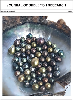
Pearls are gems produced by biological processes within the living tissues of molluscs. Image pearls are a form of blister pearl that portray a design or image such as a bird, fish, flower, goddess, etc. To achieve this, a paraffin wax mold is inserted between the shell and the mantle of a freshwater mussel and subsequent nacre coverage produces an image pearl after an appropriate culture period for the mussel. The optimum culture period has not yet been determined to maximize the quality of image pearls produced in Bangladesh, and this knowledge gap was addressed in this study. Image pearl production using the freshwater mussel Lamellidens marginalis was conducted over three culture periods (T1 = 7 mo, T2 = 9 mo, and T3 = 11 mo). A total of 3,150 mussels were implanted with 2.5 × 1.5 cm2 paraffin wax molds, with 1,050 mussels allocated to each treatment, which had five replicates. Survival and pearl production rate of mussels were negatively correlated with culture period. Survival and pearl production rate were 14.7%, 12.3%, and 11.9% for mussels cultured for 7, 9, and 11 mo, respectively. The thicknesses of the nacre making up the image pearl and pearl luster were both improved with longer culture periods. The highest quality pearls with a mean nacre thickness of 0.71 mm were produced after 11 mo. Pearls produced after 11 mo also had the highest luster (40.05 lux) compared with pearls cultured for 7 mo (12.85 lux) or 9 mo (30.2 lux). There were statistically significant differences at the P < 0.05 level in survival rate F (2, 12) = 9.40, P = 0.004 and nacre layer thickness F (2, 12) = 13.30, P = 0.001 between T1 and T3. The results indicated that image pearls with high luster and improved quality are produced after longer culture durations and confirms the influence of culture period on image pearl production and quality. Further research is required to improve image pearl production methods and pearl yield.
In general, pearl cultivation has advanced with organisms produced and improved in hatchery; however, wild spat collection is still the primary source for commercial cultivation. In Mexico, progress toward pearl oyster hatchery spat production is yet in the experimental stage. The only three farms established in the country (Guaymas, La Paz, and Cozumel Island) rely on extensive culture. Constant spat supply is imperative to ensure a successful operation; at La Paz Bay, however, larval uptake of the pearl oyster Pinctada mazatlanica in artificial collectors has always been low. This study aimed to assess spatio-temporal and bathymetric patterns of spat distribution across the area to locate suitable sites to install spat collecting stations. Our results showed significant differences (P < 0.05) between stations. Mogote, Caimancito, Punta Colorada, and Gaviota Island were the stations with the highest spat abundance at 1-m depth in July–September after 40 and 60 days of immersion. The data were also analyzed with generalized linear models. The best models, selected with the Akaike and Bayesian criteria, suggest that 1-m depth, 40- and 60-day immersion time, and site (particularly, Caimancito Beach, Punta Colorada, and Gaviota Island) contributed significantly to explaining the highest catches (mean: 5–8 organisms/collector) of P. mazatlanica spat in La Paz Bay. Therefore, the area within these sites should be considered suitable for installing the network of artificial collectors during the season of high spat abundance.
Some genera of bivalve molluscs have an iconic ancestral history that predominates in the culture of a civilization through time. The Pacific thorny oyster Spondylus crassisquama is a living example of this. This study presents the description of the early life cycle until obtaining juveniles; and, for the latter, different types of substrates were evaluated. Larval culture was developed under laboratory conditions, chronologically recording the changes in larval morphology and behavior. The thorny oyster presents the first cell cleavage at 50 min postfertilization (PF). The transition between trochophore phase and the first D-veliger larvae was observed around 20–24 h PF. Above 75% of the larvae with presence of a serrate double ring showed ocular eyespot on the 16th day PF. Between the 16th and 18th days PF, the formation of the foot of the larvae was observed. The first postlarvae were observed on the 20th day with a total length of 553.0 ± 150.10 µm, distinguished by the appearance of the dissoconch. After 30 days PF, the first settled juveniles were observed with sizes of 1.6 ± 0.41 mm. Finally, the animals showed a preference for settlement on stones and concrete, and to a lesser extent on broken shells of S. crassisquama. This study shows that juvenile production is feasible under laboratory conditions; however, this requires more research to optimize the nursery process.
The giant honeycomb oyster Hyotissa hyotis occurs at a high density in the shallow subtidal rocky bottom of Jeju Island off the south coast of Korea, where the sea surface temperature ranges from 14°C to 25°C seasonally. Unlike other oysters, H. hyotis has an extraordinarily large adductor muscle (AM) that accounts for more than 40% of the total tissue weight. This study analyzed the proximate composition, amino acids, and fatty acids of the AM of H. hyotis in Jeju Island to evaluate the nutritional potential. For the analyses, oysters were grouped based on their reproductive condition as prespawning (May–August), spawning (September–November), and postspawning (December–April). The AM contained a high level of protein (54.7%–69.4%) followed by carbohydrate (16.0%–25.3%) and lipid (4.5%–9.9%) throughout the year, indicating that the AM is enriched with protein. The total amino acids (TAA) in the AM ranged from 9.4 to 12.9 g/100 g dry weight annually, and essential amino acids accounted for up to 36.7% of the TAA. Like other marine bivalves, the AM contained a high level of taurine, which accounted for 26.5%–28.5% of the total free amino acids. The AM also included high levels of essential fatty acids, such as palmitic acid, docosahexaenoic acid, and eicosapentaenoic acid, as these fatty acids accounted for 35.9%–37.0% of the total fatty acids. The AM of prespawning oysters collected during May and August contained comparatively higher total proteins and carbohydrates. Results suggested that, like scallops and pen shells, the AM of the giant honeycomb oyster is highly suitable as seafood, and H. hyotis should be considered as a species of high potential for future aquaculture efforts.
The spatial expression patterns of three genes [kelch-like protein homolog 10 (klhl10); armadillo repeat-containing protein 4 isoform X2 (armc4); mitochondria cytochrome oxidase I (mt-co1)] known to be important mechanistic components of spermatogenesis/spermiogenesis were localized in mantle tissue sections of male Mytilus edulis using nonisotopic in situ hybridization and RNA probes based on sequences derived from available mantle RNA sequencing libraries. The expression patterns showed that all three genes are primarily localized in a layer of cells adjacent to the outer wall of the testicular acini. This layer is known to contain developing stages of spermatogonia, spermatocytes, and spermatids. Kelch-like protein homolog 10 was expressed in immature spermatogonial cells/spermatocytes characterized by large round central nuclei and situated near the periphery of the testicular acini with little to no expression in adjacent populations of spermatids or mature spermatozoa at the center of the acini. Like klhl10, armc4 transcripts were also detected in the cells near the inner periphery of the outer acinar wall. In contrast, however, the armc4 expressing cells appeared to be spatially shifted more toward the inner edge of the cell layer and toward the center of the acini. The localization of cells expressing armc4 transcripts near the inner edge of the developing sperm layer in the testicular acini of M. edulis supports a role for armc4 in spermatid morphogenesis and maturation. Mitochondria-specific cytochrome c oxidase transcripts were localized in cells throughout the inner periphery of the testicular acini wall but not in association with mature spermatozoa in the center of the acini reflecting the significant level of cellular activity that occurs during early sperm development. This supports an important mitochondrial role in early and mid-spermatogenesis in M. edulis potentially related to sperm quality control. This work provides a first view into the molecular mechanisms behind spermatogenesis/spermiogenesis in M. edulis.
Despite estimates of very high (>99.9%) pre-settlement mortality, the extremely large numbers of eggs released into the plankton means that there are abundant post-larval softshell clams (Mya arenaria) available to settle and populate intertidal flats. Many seemingly suitable flats remain devoid of clams, presumably due to post-settlement predation. Predator exclusion/clam recruitment boxes (wooden frames covered top and bottom with a fine plastic screening) were set on a mudflat in Salem Harbor, Salem, MA from April to November 2019. Clam larvae were able to settle through the screen into the boxes but large (>1.9 mm) predators such as European green crabs (Carcinus maenas) and milky ribbon worms (Cerebratulus lacteus) were excluded. At the end of the study, four of five boxes contained between 121 and 290 juvenile softshell clams ranging in size from 7 mm to 32 mm shell length. A bimodal frequency distribution of sizes likely illustrates an early summer (June) set represented by a larger size grouping followed by a second late-summer set represented by a smaller size grouping. Small green crabs (9–46 mm carapace width) found in all five boxes must have grown from extremely small stage 1 crabs that were able to settle or crawl through the screening as recently settled individuals. Resulting empty clam shells could be paired to yield the equivalent of additional clams, making the total number per box between 147 and 417 individuals. No clams were found in samples outside of the boxes, indicating that the absence of a clam population on the mudflat is due to post-settlement predation and not pre-settlement mortality.
The life history including embryonic development of most species of octopus is still poorly understood. This makes the identification of eggs and juveniles difficult, hampering distribution and dispersal studies. The Pacific pygmy octopus, Paroctopus digueti (Perrier & Rocheburne 1894), exhibits features, including its direct embryonic development, that make it an ideal candidate for aquaculture. This study provides detailed information on the embryonic development, the morphological characteristics of eggs, and the fecundity of wild female P. digueti maintained under laboratory conditions that replicate natural environmental conditions. The Pacific pygmy octopus showed a monocyclic spawning pattern that takes place in time-separated batches, leading to asynchronous embryonic development in the batch. Eggs are between 7 and 10 mm (8.9 ± 0.71 mm) in total length. During the embryonic development, 31 distinct stages were identified with a total duration of 38 days. The distribution of chromatophores showed a specific pattern, with dorsal chromatophores being more abundant and larger than ventral ones. An observed fecundity of 300 eggs per female was twice as high as the value previously reported for this species in Bahia Choya, Sonora. This study contributes to the better understanding of the life cycle of P. digueti. Besides being a basic reproductive aspect, fecundity is a key element for studies on the reproductive potential and population dynamics of the species.
Sea urchins are important inhabitants of many marine ecosystems. They are also an economic resource for many international fisheries and are an important animal model in developmental biology. Sea urchins ingest a variety of plant and animal matter, form a digesta pellet, and produce an egesta (fecal) pellet that contributes to benthic food webs. The size and morphology of fecal pellets produced by various size classes of the sea urchin Lytechinus variegatus fed a variety of natural, vegetable-based, or formulated diets were characterized in terms of two-dimensional area, length (long diameter), width (short diameter), and circularity, as well as by ultrastructure analysis using transmission electron microscopy (TEM). Sea urchins fed natural and vegetable-based ingredients produced a wide variety of irregularly shaped egesta that were loosely surrounded by mucus, whereas urchins fed a formulated diet produced highly circular egesta surrounded by a uniform multilayered mucus coat. Larger urchins (62 ± 3.1 g wet weight) consuming a formulated diet produced egesta that were larger in size than smaller adult (26.5 ± 2.7 g) or juvenile urchins (3.6 ± 0.4 g), with egesta length of 0.93 ± 0.06, 0.75 ± 0.05, and 0.61 ± 0.01 mm, respectively (mean ± 95% confidence interval [CI]; P < 0.001). Ultrastructural analysis of the egesta using TEM image analysis software from urchins consuming formulated diets indicated that pellets contained extensive bacterial communities, ranging from 31% to 69% of the total cross-sectional area of the pellet, with dense communities located near the surface of the pellet (9.51 × 10⁸ cells/cm2). Qualitative examination of the egesta TEM suggested the presence of a diverse bacterial community. Following egestion, these microbial communities are suggested to have an important role in natural food webs, with potential value for integrated multitrophic aquaculture systems.
Freshwater clams, better known as pokea clams in Indonesia, are among the important economy-generating resources whose population continues to decline in line with the increase in fishing activities. The present study aims to determine the age group, growth, mortality, and exploitation rate of pokea clams. This research was conducted in the estuary segment of Laeya River, Southeast Sulawesi, Indonesia, from March 2016 to February 2017. Pokea samples were taken using a traditional fishing tool called Tangge. Data on age groups, growth, mortality (natural, fishing, and total) and exploitation rate were processed using the Bhattacharya method, the von Bertalanffy inverse function, the width converted catch curve, and Pauly's empirical formula, respectively, accommodated in the FiSAT II program version 3.0. The results showed that the male and female pokea were spread out in 1 and 2 size groups. Male pokea was dominated by 2 size groups, whereas the female pokea was dominated by 1 size group. The growth of the male and female clams followed the equations Lt = 83.89 – (83.89–0.025)e–0.54t and Lt = 77.38 – (77.38–0.025)e–0.52t. The male natural mortality (M), fishing mortality (F), and total mortality (Z) were 2.04 y–1, 0.91 y–1, and 2.94 y–1, respectively, whereas the natural mortality, fishing mortality, and total mortality of the female clams were 1.51 y–1, 0.90 y–1, and 2.41 y–1, respectively. In general, the male and female pokea clams in the Laeya River are overexploited, with the exploitation rates of 0.69 and 0.63, respectively.
This article is only available to subscribers. It is not available for individual sale.
Access to the requested content is limited to institutions that have
purchased or subscribe to this BioOne eBook Collection. You are receiving
this notice because your organization may not have this eBook access.*
*Shibboleth/Open Athens users-please
sign in
to access your institution's subscriptions.
Additional information about institution subscriptions can be foundhere