BioOne.org will be down briefly for maintenance on 14 May 2025 between 18:00-22:00 Pacific Time US. We apologize for any inconvenience.
Registered users receive a variety of benefits including the ability to customize email alerts, create favorite journals list, and save searches.
Please note that a BioOne web account does not automatically grant access to full-text content. An institutional or society member subscription is required to view non-Open Access content.
Contact helpdesk@bioone.org with any questions.
Uncontrolled generation of DNA double-strand breaks (DSBs) in cells is regarded as a highly toxic event that threatens cell survival. Radiation-induced DNA DSBs are commonly measured by pulsed-field gel electrophoresis, microscopic evaluation of accumulating DNA damage response proteins (e.g., 53BP1 or γ-H2AX) or flow cytometric analysis of γ-H2AX. The advantage of flow cytometric analysis is that DSB formation and repair can be studied in relationship to cell cycle phase or expression of other proteins. However, γ-H2AX is not able to monitor repair kinetics within the first 60 min postirradiation, a period when most DSBs undergo repair. A key protein in non-homologous end joining repair is the catalytic subunit of DNA-dependent protein kinase. Among several phosphorylation sites of DNA-dependent protein kinase, the threonine at position 2609 (T2609), which is phosphorylated by ataxia telangiectasia mutated (ATM) or DNA-dependent protein kinase catalytic subunit itself, activates the end processing of DSB. Using flow cytometry, we show here that phosphorylation at T2609 is faster in response to DSBs than γ-H2AX. Furthermore, flow cytometric analysis of T2609 resulted in a better representation of fast repair kinetics than analysis of γ-H2AX. In cells with reduced ligase IV activity, and wild-type cells where DNA-dependent protein kinase activity was inhibited, the reduced DSB repair capacity was observed by T2609 evaluation using flow cytometry. In conclusion, flow cytometric evaluation of DNA-dependent protein kinase T2609 can be used as a marker for early DSB repair and gives a better representation of early repair events than analysis of γ-H2AX.
Radiation stimulates the expression of inflammatory mediators known to increase cancer cell invasion. Therefore, it is important to determine whether anti-inflammatory drugs can prevent this adverse effect of radiation. Since cyclooxygenase-2 (COX-2) is a central player in the inflammatory response, we performed studies to determine whether the COX-2 inhibitor NS-398 can reduce the radiation enhancement of cancer cell invasion. Thighs of Balb/c mice treated with NS-398 were irradiated with either daily fractions of 7.5 Gy for five consecutive days or a single 30 Gy dose prior to subcutaneous injection of nonirradiated MC7-L1 mammary cancer cells. Five weeks later, tumor invasion, blood vessel permeability and interstitial volumes were assessed using magnetic resonance imaging (MRI). Matrix metalloproteinase-2 (MMP-2) was measured in tissues by zymography at 21 days postirradiation. Cancer cell invasion in the mouse thighs was increased by 12-fold after fractionated irradiations (5 × 7.5 Gy) and by 17-fold after a single 30 Gy dose of radiation. This stimulation of cancer cell invasion was accompanied by a significant increase in the interstitial volume and a higher level of the protease MMP-2. NS-398 treatment largely prevented the stimulation of cancer cell invasion, which was associated with a reduction in interstitial volume in the irradiated thighs and a complete suppression of MMP-2 stimulation. In conclusion, this animal model using MC7-L1 cells demonstrates that radiation-induced cancer cell invasion can be largely prevented with the COX-2 inhibitor NS-398.
A partial-body irradiation model with approximately 2.5% bone marrow sparing (PBI/BM2.5) was established to determine the radiation dose-response relationships for the prolonged and delayed multi-organ effects of acute radiation exposure. Historically, doses reported to the entire body were assumed to be equal to the prescribed dose at some defined calculation point, and the dose-response relationship for multi-organ injury has been defined relative to the prescribed dose being delivered at this point, e.g., to a point at mid-depth at the level of the xiphoid of the non-human primate (NHP). In this retrospective-dose study, the true distribution of dose within the major organs of the NHP was evaluated, and these doses were related to that at the traditional dose-prescription point. Male rhesus macaques were exposed using the PBI/BM2.5 protocol to a prescribed dose of 10 Gy using 6-MV linear accelerator photons at a rate of 0.80 Gy/min. Point and organ doses were calculated for each NHP from computed tomography (CT) scans using heterogeneous density data. The prescribed dose of 10.0 Gy to a point at midline tissue assuming homogeneous media resulted in 10.28 Gy delivered to the prescription point when calculated using the heterogeneous CT volume of the NHP. Respective mean organ doses to the volumes of nine organs, including the heart, lung, bowel and kidney, were computed. With modern treatment planning systems, utilizing a three-dimensional reconstruction of the NHP's CT images to account for the variations in body shape and size, and using density corrections for each of the tissue types, bone, water, muscle and air, accurate determination of the differences in dose to the NHP can be achieved. Dose and volume statistics can be ascertained for any body structure or organ that has been defined using contouring tools in the planning system. Analysis of the dose delivered to critical organs relative to the total-body target dose will permit a more definitive analysis of organ-specific effects and their respective influence in multiple organ injury.
Radiation nephropathy is one of the common late effects in cancer survivors who received radiotherapy as well as in victims of radiation accidents. The clinical manifestations of radiation nephropathy occur months after exposure. To date, there are no known early biomarkers to predict the future development of radiation nephropathy. This study focuses on the development of urinary biomarkers providing readout of acute responses in renal tubular epithelial cells. An amplification-free hybridization-based nCounter assay was used to detect changes in mouse urinary miRNAs after irradiation. After a single LD50 of total-body irradiation (TBI) or clinically relevant fractionated doses (2 Gy twice daily for 3 days), changes in urinary levels of microRNAs followed either an early pattern, peaking at 6–8 h postirradiation and gradually declining, or later pattern, peaking from 24 h to 7 days. Of 600 miRNAs compared, 12 urinary miRNAs showed the acute response and seven showed the late response, common to both irradiation protocols. miR-1224 and miR-21 were of particular interest, since they were the most robust acute and late responders, respectively. The early responding miR-1224 also exhibited good dose response after 2, 4, 6 and 8 Gy TBI, indicating its potential use as a biomarker for radiation exposure. In situ hybridization of irradiated mouse kidney sections and cultured mouse primary renal tubular cells confirmed the tubular origin of miR-1224. A significant upregulation in hsa-miR-1224-3p expression was also observed in human proximal renal tubular cells after irradiation. Consistent with mouse urine data, a similar expression pattern of hsa-miR-1224-3p and hsa-miR-21 were observed in urine samples collected from human leukemia patients preconditioned with TBI. This proof-of-concept study shows the potential translational utility of urinary miRNA biomarkers for radiation damage in renal tubules with possible prediction of late effects.
Advanced radiotherapy techniques such as intensity modulated radiation therapy achieve highly conformal dose distributions within target tumor volumes through the sequential delivery of multiple spatially and temporally modulated radiation fields and have been shown to influence radiobiological response. The goals of this study were to determine the effect of hypoxia on the cell survival responses of different cell models (H460, DU145, A549, MDA231 and FADU) to modulated fields and to characterize the time dependency of signaling under oxic conditions, following reoxygenation and after prolonged hypoxia. Hypoxia was induced by incubating cells at 95% nitrogen and 5% carbon dioxide for 4 h prior to irradiation. The out-of-field response in MDA231 cells was oxygen dependent and therefore selected for co-culture studies to determine the signaling kinetics at different time intervals after irradiation under oxic and hypoxic conditions. Under both oxic and hypoxic conditions, significant increases in cell survival were observed in-field with significant decreases in survival observed out-of-field (P < 0.05), which were dependent on intercellular communication. The in-field response of MDA231 cells showed no significant time dependency up to 24 h postirradiation, while out-of-field survival decreased significantly during the first 6 h postirradiation (P < 0.05). While in-field responses were oxygen dependent, out-of-field effects were observed to be independent of oxygen, with similar or greater cell killing under hypoxic conditions. This study provides further understanding of intercellular signaling under hypoxic conditions and highlights the need for further refinement of established radiobiological models for future applications in advanced radiotherapies.
In this study, lung cancer risk from occupational plutonium exposure was analyzed in a pooled cohort of Mayak and Sellafield workers, two of the most informative cohorts in the world with detailed plutonium urine monitoring programs. The pooled cohort comprised 45,817 workers: 23,443 Sellafield workers first employed during 1947–2002 with follow-up until the end of 2005 and 22,374 Mayak workers first employed during 1948–1982 with follow-up until the end of 2008. In the pooled cohort 1,195 lung cancer deaths were observed (789 Mayak, 406 Sellafield) but only 893 lung cancer incidences (509 Mayak, 384 Sellafield, due to truncated follow-up in the incidence analysis). Analyses were performed using Poisson regression models, and were based on doses derived from individual radiation monitoring data using an updated dose assessment methodology developed in the study. There was clear evidence of a linear association between cumulative internal plutonium lung dose and risk of both lung cancer mortality and incidence in the pooled cohort. The pooled point estimates of the excess relative risk (ERR) from plutonium exposure for both lung cancer mortality and incidence were within the range of 5–8 per Gy for males at age 60. The ERR estimates in relationship to external gamma radiation were also significantly raised and in the range 0.2–0.4 per Gy of cumulative gamma dose to the lung. The point estimates of risk, for both external and plutonium exposure, were comparable between the cohorts, which suggests that the pooling of these data was valid. The results support point estimates of relative biological effectiveness (RBE) in the range of 10–25, which is in broad agreement with the value of 20 currently adopted in radiological protection as the radiation weighting factor for alpha particles, however, the uncertainty on this value (RBE = 21; 95% CI: 9–178) is large. The results provide direct evidence that the plutonium risks in each cohort are of the same order of magnitude but the uncertainty on the Sellafield cohort plutonium risk estimates is large, with observed risks consistent with no plutonium risk, and risks five times larger than those observed in the Mayak cohort.
The development of medical countermeasures against acute and delayed multi-organ injury requires animal models predictive of the human response to radiation and its treatment. Late chronic injury is a well-known feature of radiation nephropathy, but acute kidney injury has not been reported in an appropriate animal model. We have established a single-fraction partial-body irradiation model with minimal marrow sparing in non-human primates. Subject-based medical management was used including parenteral fluids according to prospective morbidity criteria. We show herein that 10 or 11 Gy exposures caused both acute and chronic kidney injury. Acute and chronic kidney injury appear to be dose-independent between 10 and 11 Gy. Acute kidney injury was identified during the first 50 days postirradiation and appeared to resolve before the occurrence of chronic kidney injury, which was progressively more severe up to 180 days postirradiation, which was the end of the study. These findings show that mitigation of the acute radiation syndrome by medical management will unmask delayed late effects that occur months after partial-body irradiation. They further emphasize that both acute and chronic changes in kidney function must be taken into account in the use and timing of mitigators and medical management for acute radiation syndrome and delayed effects of acute radiation exposure (DEARE).
Efforts to protect astronauts from harmful galactic cosmic radiation (GCR) require a better understanding of the effects of GCR on human health. In particular, little is known about the lasting effects of GCR on the central nervous system (CNS), which may lead to behavior performance deficits. Previous studies have shown that high-linear energy transfer (LET) radiation in rodents leads to short-term declines in a variety of behavior tests. However, the lasting impact of low-, medium- and high-LET radiation on behavior are not fully defined. Therefore, in this study C57BL/6 male mice were irradiated with 100 or 250 cGy of γ rays (LET ∼0.3 KeV/μm), 10 or 100 cGy of 1H at 1,000 MeV/n (LET ∼0.2 KeV/μm), 28Si at 300 MeV/n (LET ∼69 KeV/μm) or 56Fe at 600 MeV/n (LET of ∼180 KeV/μm), and behavior metrics were collected at 5 and 9 months postirradiation to analyze differences among radiation qualities and doses. A significant dose effect was observed on recognition memory and activity levels measured 9 months postirradiation, regardless of radiation source. In contrast, we observed that each ion species had a distinct effect on anxiety, motor coordination and spatial memory at extended time points. Although 28Si and 56Fe are both regarded as high-LET particles, they were shown to have different detrimental effects on behavior. In summary, our findings suggest that GCR not only affects the CNS in the short term, but also has lasting damaging effects on the CNS that can cause sustained declines in behavior performance.
Transit amplifying cells (TACs) are highly proliferative in nature and tend to be sensitive to ionizing radiation. Due to the abundance of TACs that support the elongation of hair shafts, growing hair follicles are highly sensitive to radiation injury. How hair follicles repair themselves after radiation injury is unclear. In this study, we observed that in 4 Gy irradiated mice, hair follicle dystrophy was induced with apoptosis-driven loss of hair matrix cells, which are the TACs that fuel hair growth. The dystrophy was repaired within 96 h without significant hair loss, indicating that a regenerative attempt successfully restored the TAC population to resume anagen growth. Soon after irradiation, mTORC1 signaling was activated in the TAC compartment and its activation was maintained until the regeneration process was completed. Inhibition of mTORC1 by rapamycin treatment increased radiation-induced cell apoptosis, reduced cell proliferation and delayed restoration of Wnt signaling in the hair matrix after radiation injury, leading to prolonged dystrophy and hair loss. These results demonstrate that mTORC1 signaling is activated after irradiation and is required for timely regeneration of the TAC pool of hair follicles, so that hair growth can resume after radiation injury.
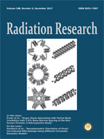
Monte Carlo based simulation has proven useful in investigating the effect of proton-induced DNA damage and the processes through which this damage occurs. Clustering of ionizations within a small volume can be related to DNA damage through the principles of nanodosimetry. For simulation, it is standard to construct a small volume of water and determine spatial clusters. More recently, realistic DNA geometries have been used, tracking energy depositions within DNA backbone volumes. Traditionally a chromatin fiber is built within the simulation and identically replicated throughout a cell nucleus, representing the cell in interphase. However, the in vivo geometry of the chromatin fiber is still unknown within the literature, with many proposed models. In this work, the Geant4-DNA toolkit was used to build three chromatin models: the solenoid, zig-zag and cross-linked geometries. All fibers were built to the same chromatin density of 4.2 nucleosomes/11 nm. The fibers were then irradiated with protons (LET 5–80 keV/μm) or alpha particles (LET 63–226 keV/μm). Nanodosimetric parameters were scored for each fiber after each LET and used as a comparator among the models. Statistically significant differences were observed in the double-strand break backbone size distributions among the models, although nonsignificant differences were noted among the nanodosimetric parameters. From the data presented in this article, we conclude that selection of the solenoid, zig-zag or cross-linked chromatin model does not significantly affect the calculated nanodosimetric parameters. This allows for a simulation-based cell model to make use of any of these chromatin models for the scoring of direct ion-induced DNA damage.
This article is only available to subscribers. It is not available for individual sale.
Access to the requested content is limited to institutions that have
purchased or subscribe to this BioOne eBook Collection. You are receiving
this notice because your organization may not have this eBook access.*
*Shibboleth/Open Athens users-please
sign in
to access your institution's subscriptions.
Additional information about institution subscriptions can be foundhere