Registered users receive a variety of benefits including the ability to customize email alerts, create favorite journals list, and save searches.
Please note that a BioOne web account does not automatically grant access to full-text content. An institutional or society member subscription is required to view non-Open Access content.
Contact helpdesk@bioone.org with any questions.
Cell line misidentification and contamination are major contributors to the reproducibility crisis in academic research. Authentication of cell lines provides assurances of the data generated; however, commercially available cells are often not subjected to rigorous identification testing. In this study, commercially available cell lines underwent testing to confirm cell identity and purity. The methods reported here outline the best practices for cell line authentication. Briefly, a commercially available primary rabbit aortic endothelial cell line was purchased for the intent of producing target proteins necessary for generating species-specific recombinant antibodies. These rabbit-specific antibodies would then be utilized for the development of in-house enzyme-linked immunosorbent assays (ELISA) to evaluate blood-based biomarkers of vascular injury after total-body irradiation. To authenticate the cell line, cell identity and purity were determined by single tandem repeat (STR) testing, flow cytometry, polymerase chain reaction (PCR), and cytochrome c oxidase subunit 1 (CO1) DNA Barcoding in-house and/or through commercial vendors. Fresh cells obtained from a New Zealand White rabbit (Charles River, Wilmington, DE) were used as a positive control. The results of STR and flow cytometry analyses indicated the cells were not contaminated with human or mouse cells, and that the cells were not of endothelial origin. PCR demonstrated that cells were also not of rabbit origin, which was further confirmed by a third-party vendor. An unopened vial of cells was submitted to another vendor for CO1 DNA Barcoding analysis, which identified the cells as being purely of bovine origin. Results revealed that despite purchase through a commercial vendor, the cell line marketed as primary rabbit aortic endothelial cells were of bovine origin. Purity analysis found cells were misidentified rather than contaminated. Further investigation to determine the cell type was not performed. The most cost-effective and efficient methodology for confirming cell line identity was found to be CO1 DNA Barcoding performed by a commercial vendor.
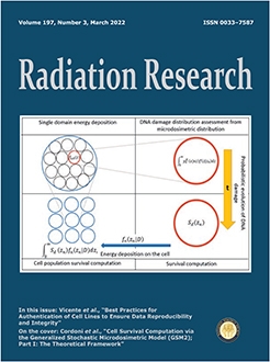
The current article presents the first application of the Generalized Stochastic Microdosimetric Model (GSM2) for computing explicitly a cell survival curve. GSM2 is a general probabilistic model that predicts the kinetic evolution of DNA damages taking full advantage of a microdosimetric description of a radiation energy deposition. We show that, despite the high generality and flexibility of GSM2, an explicit form for the survival fraction curve predicted by the GSM2 is achievable. We illustrate how several correction terms typically added a posteriori in existing radiobiological models to improve the prediction accuracy, are naturally included into GSM2. Among the most relevant features of the survival curve derived from GSM2 and presented in this article, is the linear-quadratic behavior at low doses and a purely linear trend for high doses. The study also identifies and discusses the connections between GSM2 and existing cell survival models, such as the Microdosimetric Kinetic Model (MKM) and the Multi-hit model. Several approximations to predict cell survival in different irradiation regimes are also introduced to include intercellular non-Poissonian behaviors.
Jean Albert Laissue, Sébastien Barré, Stefan Bartzsch, Hans Blattmann, Audrey M. Bouchet, Valentin G. Djonov, David Haberthür, Ruslan Hlushchuk, Barbara Kaser-Hotz, Pierre Philippe Laissue, Géraldine LeDuc, Susanne Oswald Reding, Raphaël Serduc
Microbeam radiation therapy, an alternative radiosurgical treatment under preclinical investigation, aims to safely treat muzzle tumors in pet animals. This will require data on the largely unknown radiation toxicity of microbeam arrays for bones and teeth. To this end, the muzzle of six young adult New Zealand rabbits was irradiated by a lateral array of microplanar beamlets with peak entrance doses of 200, 330 or 500 Gy. The muzzles were examined 431 days postirradiation by computed microtomographic imaging (micro-CT) ex vivo, and extensive histopathology. The boundaries of the radiation field were identified histologically by microbeam tracks in cartilage and other tissues. There was no radionecrosis of facial bones in any rabbit. Conversely, normal incisor teeth exposed to peak entrance doses of 330 Gy or 500 Gy developed marked caries-like damage, whereas the incisors of the two rabbits exposed to 200 Gy remained unscathed. A single, unidirectional array of microbeams with a peak entrance dose ≤200 Gy (valley dose14 Gy) did not damage normal bone, teeth and soft tissues of the muzzle of normal rabbits longer than one year after irradiation. Because of that, Microbeam radiation therapy of muzzle tumors in pet animals is unlikely to cause sizeable damage to normal teeth, bone and soft tissues, if a single array as used here delivers a limited entrance dose of 200 Gy and a valley dose of ≤14 Gy.
Beyond low-Earth orbit, space radiation poses significant risks to astronaut health. Previous studies have shown that the microbial composition of the gastrointestinal (GI) microbiome changes upon exposure to high-linear energy transfer radiation. Interestingly, radiation-induced shifts in GI microbiota composition are linked to various neuropsychological disorders. Herein, we aimed to study changes in GI microbiota and behaviors of rats exposed to whole-body radiation (0, 5 or 25 cGy 4He, 250 MeV/n) at approximately 6 months of age. Fecal samples were collected 24 h prior to 4He irradiation and 24 h and 7 days postirradiation for quantitative PCR analyses to assess fecal levels of spore-forming bacteria (SFB), Bifidobacterium, Lactobacillus and Akkermansia. Rats were also tested in the social odor recognition memory (SORM) test at day 7 after 4He exposure. A subset of rats was euthanized 90 min after completion of the SORM test, and GI tissue from small intestine to colon were prepared for examining overall histological changes and immunohistochemical staining for serotonin (5-HT). No notable pathological changes were observed in GI tissues. Akkermansia spp. and SFB were significantly decreased in the 25 cGy group at 24 h and 7 days postirradiation compared to pre-exposure, respectively. Bifidobacterium and Lactobacillus spp. showed no significant changes. 5-HT production was significantly higher in the proximal small intestine and the cecum in the 25 cGy group compared to the sham group. The 25 cGy group exhibited deficits in recognition in SORM testing at day 7 postirradiation. Taken together, these results suggest a connection between GI microbiome composition, serotonin production, and neurobehavioral performance, and that this connection may be disrupted upon exposure to 25 cGy of 4He ions.
Charles A. Maitz, Deborah Tate, Sandra Bechtel, Joni Lunceford, Carolyn Henry, Brian Flesner, Amanda Collins, Mary Varterasian, David Tung, Linping Zhang, Saurabh Saha, Jeffrey N. Bryan
Hypoxia is associated with neoplastic tissue, protecting cancer cells from death by irradiation and chemotherapy. Identification of hypoxic volume of tumors could optimize patient selection for hypoxia-directed medical, immunological, and radiation therapies. Clostridium novyi-NT (CNV-NT) is an oncolytic bacterium derived from attenuated wild-type Clostridium novyi spores, which germinates exclusively in the anaerobic core of tumors with low-oxygen content. The hypothesis was that 64Cu-ATSM would localize to regions of hypoxia, and that greater hypoxic volume would result in greater germination of Clostridium novyi-NT (CNV-NT). Tumor-bearing companion dogs were recruited to a veterinary clinical trial. Dogs received a CT scan, 18F-FDG PET scan (74 MBq) and 64Cu-ATSM PET scan (74 MBq). Scan regions of interest were defined as the highest 20% of counts/voxel for each PET scan, and regions with voxels overlapping between the two scans. Maximum standardized uptake value (MaxSUV) and threshold volume were calculated. Direct oximetry was performed in select tumors. Tumor types evaluated included nerve sheath tumor (10), apocrine carcinoma (1), melanoma (3) and oral sarcoma (6). MaxSUVATSM ranged from 0.3–6.6. Measured oxygen tension ranged from 0.05–89.9 mmHg. Inverse of MaxSUVATSM had a linear relationship with oxygen tension (R2 = 0.53, P = 0.0048). Hypoxia <8 mmHg was associated with an SUVATSM > 1.0. Hypoxic volume ranged from 0 to 100% of gross tumor volume (GTV) and MaxSUVATSM was positively correlated with hypoxic volume (R = 0.674; P = 0.0001), but not GTV (P = 0.182). Tumor hypoxic volume was heterogeneous in location and distribution. 64Cu-ATSM-avid regions were associated with differential CT attenuation. Hypoxic volume did not predict CNV-NT germination. 64Cu-ATSM PET scanning predicts hypoxia patterns within spontaneously occurring tumors of dogs as measured by direct oxymetry. Total tumor volume does not accurately predict degree or proportion of tumor hypoxia.
Metaphase-based cytogenetic methods based on scoring of chromosome aberrations for the estimation of the radiation dose received provide a powerful approach for evaluating the associated risk upon radiation exposure and form the bulk of our current knowledge of radiation-induced chromosome damages. They mainly rely on inducing quiescent peripheral lymphocytes into proliferation and blocking them at metaphases to quantify the damages at the chromosome level. However, human organs and tissues demonstrate various sensitivity towards radiation and within them, self-proliferating progenitor/stem cells are believed to be the most sensitive populations. The radiation-induced chromosome aberrations in these cells remain largely unknown, especially in the context of an intact living organism. Zebrafish is an ideal animal model for research into this aspect due to their small size and the large quantities of progenitor cells present during the embryonic stages. In this study, we employ a novel metaphase-based cytogenetic approach on zebrafish embryos and demonstrate that chromosome-type and chromatid-type aberrations could be identified in progenitor cells at different cell-cycle stages at the point of radiation exposure. Our work positions zebrafish at the forefront as a useful animal model for studying radiation-induced chromosome structural changes in vivo.
Marina A. Yakovleva, Tatiana B. Feldman, Kristina N. Lyakhova, Dina M. Utina, Inna A. Kolesnikova, Yuliya V. Vinogradova, Alexander G. Molokanov, Mikhail A. Ostrovsky
The present study evaluated the effects of proton and gamma-ray ionizing radiation on the mouse eye. The aim of this work was to analyze radiation-mediated retinoid oxidation in the retina and retinal pigment epithelium (RPE). The findings from this analysis can be used to develop a noninvasive method for rapid assessment of the effects of ionizing radiation. Comparative fluorescence and chromatographic analyses of retinoids before and after irradiations were performed. The fluorescent properties of chloroform extracts from irradiated mouse retina and RPE exhibited an increase in fluorescence intensity in the short-wave region of the spectrum (λ < 550 nm). This change is due to increased retinal and RPE retinoid oxidation and degradation products after radiation exposure. Comparative analyses of radiation effects demonstrated that the effect of proton exposure on the retina and RPE was higher than that of gamma-ray exposure. The present study revealed a new approach to assessing the level of radiation exposure in ocular tissues.
Several studies have reported differences in radiation toxicity between the sexes, but these differences have not been tested with respect to histopathology and genes. This animal study aimed to show an association between histopathological findings of radiation-induced lung toxicity and the genes ATM, SOD2, TGF-β1, XRCC1, XRCC3 and HHR2. In all, 120 animals were randomly divided into 2 control groups (male and female) and experimental groups comprising fifteen rats stratified by sex, radiotherapy (0 Gy vs. 10 Gy), and time to sacrifice (6, 12, and 24 weeks postirradiation). Histopathological evaluations for lung injury, namely, intra-alveolar edema, alveolar neutrophils, intra-alveolar erythrocytes, activated macrophages, intra-alveolar fibrosis, hyaline arteriosclerosis, and collapse were performed under a light microscope using a grid system; the evaluations were semi quantitatively scored. Then, the alveolar wall thickness was measured. Real-time quantitative reverse transcription PCR (RT-qPCR) was used to determine gene expression differences in ATM, TGF-β1, XRCC1, XRCC3, SOD2 and HHR2L among the groups. Histopathological data showed that radiation-induced acute, subacute, and chronic lung toxicity were worse in male rats. The expression levels of the evaluated genes were significantly higher in females than males in the control group, but this difference was lost over time after radiotherapy. Less toxicity in females may be attributable to the fact that the expression of the evaluated genes was higher in normal lung tissue in females than in males and the changes in gene expression patterns in the postradiotherapy period played a protective role in females. Additional data related to pulmonary function, lung weights, imaging, or outcomes are needed to support this data that is based on histopathology alone.
This study has established the impact that space radiation exposure has on the capability of rats to successfully negotiate behavioral tasks of increasing complexity. Rats previously exposed to a low dose (10 cGy) of either 4He ions or a cocktail of 6 ions that simulates the galactic cosmic ray spectrum (GCRSim) were screened initially on an attentional set shifting (ATSET) task that provides a measure of executive function. Rats that exhibited superior ATSET performance were then selected for follow up behavioral assessments designed to evaluate how the cohort of “good performers” would fare when presented with a novel behavioral paradigm termed the Associative Recognition Memory and Interference Touchscreen (ARMIT) task. Central to this approach was to discriminate if/how adaptive problem solving would be impacted by changing the options of associative cues presented over several learning sessions to obtain a reward under time constraints using this newly designed touch screen-based task. Data from these studies indicated that when faced with an increased cognitive load, possibly due to interference from prior associative recognition memories, rats exhibited impairments in their capability to negotiate task dynamics and efficiently engage abstract reasoning. Interestingly, while exposure to the GCRSim adversely impacted problem-solving capabilities, single ion exposure did not, pointing to the nuances of space radiation exposure on CNS functionality. Since the selected behavioral paradigms exhibit strong cross-species correlates, data suggest that rodents succumb to increased task rigor as observed in humans, and make similar mistakes when challenged with the interference of overlapping associative memories. Furthermore, data clearly points to the limitations of over-reliance on a single cognitive endpoint that may underestimate global neurocognitive risk due to space radiation exposure.
We report on effects of low-dose exposures of accelerated protons delivered at high-dose rate (HDR) or a simulated solar-particle event (SPE) like low-dose rate (LDR) on immediate DNA damage induction and processing, survival and in vitro transformation of low passage NFF28 apparently normal primary human fibroblasts. Cultures were exposed to 50, 100 and 1,000 MeV monoenergetic protons in the Bragg entrance/plateau region and cesium-137 γ rays at 20 Gy/h (HDR) or 1 Gy/h (LDR). DNA double-strand breaks (DSB) and clustered DNA damages (containing oxypurines and abasic sites) were measured using transverse alternating gel electrophoresis (TAFE) and immunocytochemical detection/scoring of colocalized γ-H2AX pS139/53BP1 foci, with their induction being linear energy transfer (LET) dependent and dose-rate sparing observed for the different damage classes. Relative biological effectiveness (RBE) values for cell survival after proton irradiation at both dose-rates ranged from 0.61–0.73. Transformation RBE values were dose-rate dependent, ranging from ∼1.8–3.1 and ∼0.6–1.0 at low doses (≤30 cGy) for HDR and LDR irradiations, respectively. However peak transformation frequencies were significantly higher (1.3–7.3-fold) for higher doses of 0.5–1 Gy delivered at SPE-like LDR. Cell survival and transformation frequencies measured after low-dose 500 MeV/n He-4, 290 MeV/n C-12 and 600 MeV/n Si-28 ion irradiations also showed an inverse dose-rate effect for transformation at SPE-like LDR. This work demonstrates the existence of inverse dose-rate effects for proton and light-ion-induced postirradiation cell survival and in vitro transformation for space mission-relevant doses and dose rates.
This article is only available to subscribers. It is not available for individual sale.
Access to the requested content is limited to institutions that have
purchased or subscribe to this BioOne eBook Collection. You are receiving
this notice because your organization may not have this eBook access.*
*Shibboleth/Open Athens users-please
sign in
to access your institution's subscriptions.
Additional information about institution subscriptions can be foundhere